

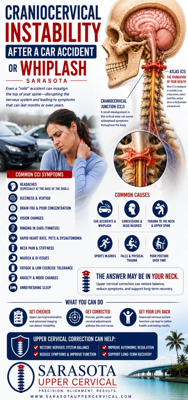
Craniocervical Instability After a Car Accident or Whiplash
Most of what is written about craniocervical instability is written for people with Ehlers-Danlos syndrome. That is reasonable — connective tissue disorders are the best-documented route to ligamentous insufficiency at the top of the neck, and the research base is concentrated there.
Schedule Your Appointment
Schedule appointmentBut it leaves out the other route, and the other route affects more people.
Ligaments do not only fail because they were built differently. They also fail because something tore them. A rear-end collision, a fall down stairs, a hard tackle, a bicycle accident, a blow to the head — any of these can load the craniocervical junction beyond what its restraints tolerate. And when that happens, the person who develops instability afterward has no connective tissue diagnosis, does not fit the profile anyone is screening for, and typically spends years being told their imaging is clear.
This article is about that group.
What the injury actually does at the top of the neck
The mechanics deserve to be specific, because the usual description — the neck snaps forward and back — is not what happens and obscures why the upper cervical spine in particular is vulnerable.
In a rear-end collision, the seat drives your torso forward while your head, weighing ten to twelve pounds and sitting atop a narrow column, initially stays where it was. Within the first hundred milliseconds, before any voluntary muscular response is possible, the cervical spine passes through an S-shaped deformation: the lower segments extend while the upper segments are still flexing. This is not a simple arc. Different levels of the neck are moving in opposite directions simultaneously, and the junction between them absorbs shear rather than smooth bending.
The craniocervical junction is where that shear concentrates, and it is the region least equipped to handle it. Everywhere else in the spine, interlocking facet joints and bony architecture limit motion mechanically. At the occiput-C1 and C1-C2 levels, that architecture is largely absent by design — traded away for the range of motion that lets you turn your head. What remains is ligament.
The transverse ligament holds the odontoid process of C2 against the anterior arch of C1, preventing forward translation of the atlas. The alar ligaments run from the odontoid to the occipital condyles, checking rotation and lateral bending. The tectorial membrane adds posterior restraint. These are the structures under load during the injury, and unlike large ligaments elsewhere they have limited redundancy — there is no second system to take over if they are compromised.

Rotation at the moment of impact worsens all of this. The alar ligaments are already partially wound and shortened when the head is turned, which reduces their available reserve. A collision taken while looking over your shoulder — checking a mirror, glancing at a passenger — loads these tissues from a mechanically disadvantaged starting position. This is why two people in the same crash can have very different outcomes.
Why nobody found it
The imaging you received after the accident was almost certainly designed to exclude the things that kill people quickly. Fracture, dislocation, arterial dissection, intracranial bleeding. That is entirely appropriate — those are the emergencies, and ruling them out is what an emergency department is for.
But a CT or X-ray in a cervical collar, or a supine MRI performed days later, is close to the ideal set of conditions for making an unstable joint look stable. The gravitational load is off. The head is supported. The neck is in neutral. Ligamentous insufficiency becomes visible under load and at end range, and none of that is present in the images that were taken.
So the report reads normal, which it accurately is with respect to the question asked, and the file closes. The specialists who evaluate this condition use upright, weight-bearing imaging captured in flexion and extension — Henderson and colleagues obtained upright weight-bearing flexion and extension MRI in half their surgical cohort in their long-term follow-up work, and Nicholson and colleagues established reference values for craniocervical stability measures using upright dynamic MRI captured at maximal flexion, maximal extension and neutral. Those are not the studies performed after a car accident.
There is also a timing problem. Acute soft tissue injury produces pain, spasm and guarding that dominate the picture for weeks or months. The muscles splint the joint, and while they are splinting it the instability is partially masked. As the acute injury settles and the guarding relaxes, the underlying insufficiency becomes more apparent — which is to say, symptoms can emerge or worsen months after the accident, at exactly the point when everyone has concluded you recovered. Patients describing this pattern are frequently disbelieved, and the mechanism is straightforward.
The symptom pattern in traumatic cases
Largely the same as instability from any cause, with a few features that recur specifically in post-traumatic presentations.
The headache is occipital or suboccipital, feels like pressure or weight rather than throbbing, and worsens the longer you stay upright or the longer you spend looking down. Lying flat helps. Many patients work this out themselves long before anyone examines them.
The heavy head sensation is characteristic and diagnostically useful. Patients describe supporting the head with a hand, propping the chin, feeling that the neck is working constantly just to hold position. It appears in few other conditions.
Symptoms provoked by head rotation are common where alar ligament involvement is likely — dizziness or symptom flare specifically on turning, often worse to one side than the other.
Cranial nerve symptoms cluster: swallowing difficulty, tinnitus, voice changes, visual disturbance, altered facial sensation. Any one alone means little. Several together, alongside the positional headache, is a pattern.
Autonomic symptoms are common and frequently the most disabling part. Racing heart on standing, lightheadedness, presyncope, exercise intolerance, temperature dysregulation. The overlap with POTS is substantial, and post-traumatic POTS and post-traumatic instability are not mutually exclusive — they may be two descriptions of consequences from the same injury.
Cognitive symptoms — fog, word-finding difficulty, poor concentration, mental fatigue that tracks with upright time — are routinely attributed to post-concussion syndrome and left there. That attribution may be correct. It may also be incomplete, particularly where symptoms are clearly positional.
Progressive neurological signs change the urgency entirely. Weakness, numbness, hand clumsiness, gait disturbance, or bowel or bladder changes suggest possible cord or brainstem involvement and require prompt medical evaluation rather than conservative care of any kind.
The medico-legal complication
Worth naming, because it affects how these patients are treated.
When symptoms follow a motor vehicle collision, an insurance claim frequently exists, and the presence of that claim changes how clinicians receive the history. Persistent symptoms after an accident with normal imaging are, in some settings, treated as presumptively exaggerated. This is a documented bias and it does real harm to people who have a genuine structural problem that the imaging was not designed to detect.
It is also true that symptom reporting in litigation contexts is genuinely complicated, and pretending otherwise would be naive. The useful response for a patient is to be precise rather than emphatic: document the mechanism of injury including head position at impact, document the timeline of symptom onset and change, note specifically what makes symptoms better and worse, and record what imaging was performed in what position. Precise, positional, mechanically coherent history is harder to dismiss than a list of complaints.
What to do
Related article
Upper Cervical Chiropractic for Migraine Relief: What Patients Should Know
Oct 20, 2025
Establish what has actually been imaged. Get the reports. Note the position — supine or upright, neutral or dynamic. If everything was supine and neutral, instability has not been assessed, whatever the reports say.
Ask for the right study from the right person. Upright dynamic MRI with flexion and extension views, read by someone who measures craniocervical parameters. The measures used in the literature include the clivo-axial angle, the Harris measurements, the Grabb-Mapstone-Oakes measurement, and the atlanto-dental interval for atlantoaxial translation. A referral generally needs to come from a neurologist or neurosurgeon familiar with the condition.
Keep the differential open. Post-traumatic headache, post-concussion syndrome, cervicogenic headache, cervical facet injury, vestibular dysfunction, and post-traumatic POTS all follow the same accidents and produce overlapping pictures. Several may be present simultaneously. Instability is one possibility among several, and pursuing it exclusively is its own kind of error.
Escalate red flags immediately. Progressive neurological signs need urgent medical assessment regardless of previous imaging.
Where upper cervical care fits — and where it does not
This requires care, and I would rather be clear than persuasive.
Where conservative management does have a role, the literature points toward stabilization rather than mobilization. Russek and colleagues published work in Frontiers in Medicine in 2023 on the presentation and physical therapy management of upper cervical instability in symptomatic generalized joint hypermobility, and the principle running through that work is retraining deep cervical stabilizers and improving proprioceptive control — building active support for a joint whose passive support is compromised. The instinct to loosen a stiff neck is frequently wrong in these patients, because the stiffness is often protective.
For upper cervical chiropractic specifically, the appropriate approach in this population is low-force and precision-based rather than manipulative, and it should follow an evaluation that has actively considered instability rather than assumed its absence. Some post-traumatic patients are reasonable candidates for that care. Others should be referred for neurological or neurosurgical evaluation instead. Distinguishing between those groups is the most important thing a clinician does here.
What I would say to anyone whose symptoms started with a crash and who has been told repeatedly that their scans are clear: the scans probably were clear, for what they were looking at. That is a narrower statement than it sounded like at the time, and there are more specific questions still worth asking.
If your symptoms began after a car accident, fall, or head injury and you want a structural evaluation of your upper cervical spine — including an honest assessment of whether conservative care is appropriate or whether you need medical referral — schedule a free consultation or call 941-259-1891.
Medical disclaimer: This article is for educational purposes only and does not constitute medical advice, diagnosis, or treatment. Craniocervical and atlantoaxial instability are medical diagnoses requiring evaluation by qualified physicians using appropriate dynamic imaging, and cervical manipulation may be contraindicated in their presence. Anyone with suspected instability should obtain medical evaluation before undertaking hands-on cervical treatment. Progressive neurological symptoms require prompt medical evaluation. Upper cervical chiropractic care is a complementary approach and is not a substitute for medical or surgical management. Individual results vary. Always consult your physician before making changes to your care.


Leave a comment