

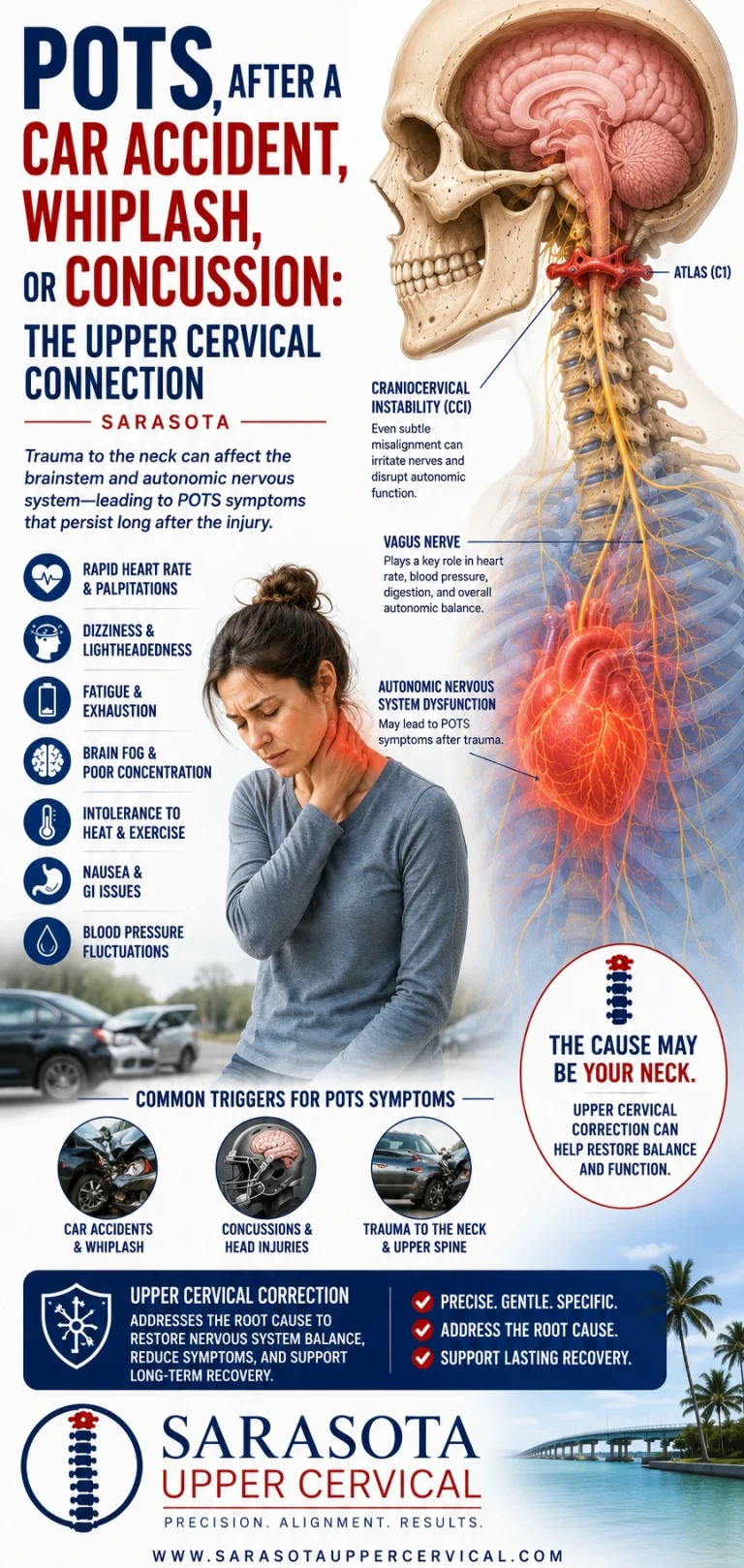
POTS After a Car Accident, Whiplash, or Concussion: The Upper Cervical Connection
There is a particular kind of POTS story that comes up again and again in our Sarasota office. The person was healthy. They were working, exercising, driving, living a normal life. Then there was a crash, or a fall, or a hit during a game. Maybe they were told they had a concussion, or maybe they were told nothing was broken and sent home with instructions to rest. The headaches and neck pain eventually settled down, more or less. But something else never came back.
Schedule Your Appointment
Schedule appointmentStanding became difficult. The heart began racing on the way up from a chair. Showers became exhausting. Grocery stores became a problem. Months later, sometimes years later, someone finally ran a tilt table test and used the words postural orthostatic tachycardia syndrome. And the question that person has almost always been carrying since the beginning is the one nobody has been able to answer: what does the accident have to do with this?
That question deserves a real answer, and it happens to be the version of POTS where the upper cervical spine has the most explanatory power. Not because upper cervical care is a cure — it is not, and this article is going to be careful about that — but because when autonomic dysfunction begins immediately after a violent loading event at the top of the neck, the neck stops being a speculative variable and becomes the most obvious thing in the room that nobody examined.
Why the timing matters more than anything else
Most POTS cases have no clear starting line. Symptoms accumulate. People look back and cannot say when it began. That ambiguity is part of why the condition takes years to diagnose and why patients are so often told their symptoms are anxiety.
Post-traumatic POTS is different. There is a date. There is an event. There is a before and an after, and the person can usually name both. That temporal relationship is not proof of causation — plenty of things happen after accidents that the accident did not cause — but in clinical reasoning, a sharp onset following a specific mechanical event is a meaningful signal, and it narrows the field considerably.
The published literature has taken notice. Kanjwal and colleagues described a group of adults who developed orthostatic intolerance following traumatic brain injury, with the interval between injury and tilt table diagnosis of POTS ranging from three months to three years. Heyer and colleagues, studying youth with persistent post-concussion symptoms, found a high rate of tilt table abnormalities and, notably, observed that in several patients the tilt table findings normalized as post-concussion symptoms improved — suggesting the autonomic disturbance was tracking with the injury rather than existing independently of it. Reviews of exercise intolerance after concussion have gone further, recommending that routine orthostatic testing be performed after both concussion and whiplash-type deceleration injuries, on the grounds that POTS is being missed in this population and may itself be a reason some people fail to recover on the expected timeline.
It is worth being straight about the fact that this evidence is not uniform. A 2025 prospective controlled study of children aged eight to eighteen compared those with persisting post-concussion symptoms against asymptomatic injured children and healthy controls, and found no significant difference in orthostatic response and no increased prevalence of POTS in the symptomatic group. The authors had hypothesized they would find one, and they did not. That result belongs in this discussion. The honest summary is that autonomic dysfunction after head and neck trauma is well recognized, that a subset of people clearly develop persistent orthostatic intolerance afterward, and that researchers are still working out how large that subset is and who is in it.
What actually happens to the upper neck in these injuries
To understand why the top of the neck comes into this conversation, it helps to be specific about what a deceleration injury does mechanically.
In a rear-end collision, the torso is driven forward by the seat while the head, which weighs roughly ten to twelve pounds and sits on top of a narrow column, lags behind and then whips forward. The resulting motion is not a simple flexion and extension arc. In the first hundred milliseconds, before any voluntary muscle response is possible, the cervical spine passes through an S-shaped deformation in which the lower segments extend while the upper segments are still flexing. The junction between skull, atlas, and axis experiences shear and rotational loading that it was never designed to absorb.
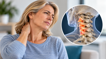
The structures that stabilize that junction are small and specific. The transverse ligament holds the odontoid process of C2 against the anterior arch of C1, preventing the atlas from translating forward on the axis. The alar ligaments run from the odontoid up to the occipital condyles and check rotation and side bending. These are the primary passive restraints at the craniocervical junction, and unlike the large ligaments lower in the spine they have limited redundancy. Partial strain to these tissues does not necessarily show up on a standard X-ray or a supine MRI, because both of those images are taken in a neutral, unloaded position — which is precisely the position in which a subtly unstable joint looks normal.
The muscular layer matters just as much. The suboccipital muscles — rectus capitis posterior major and minor, obliquus capitis superior and inferior — are unusual tissue. They are short, they cross only one or two joints, and they contain one of the highest densities of muscle spindles found anywhere in the human body. Spindle density in these muscles is orders of magnitude greater than in large limb muscles. These are not primarily muscles for producing movement. They are sensory organs that happen to be able to contract, and their job is to tell the brain, continuously and with very high resolution, exactly where the head is sitting relative to the body.
Rectus capitis posterior minor has one further feature worth knowing about: it connects to the spinal dura by a dense connective tissue structure known as the myodural bridge. There is a direct mechanical link between a suboccipital muscle and the covering of the central nervous system.
When these tissues are torn, strained, or left in protective spasm after an injury, the mechanical result may be modest. The sensory result is not.
The neurology: why degraded neck input can destabilize standing
Related article
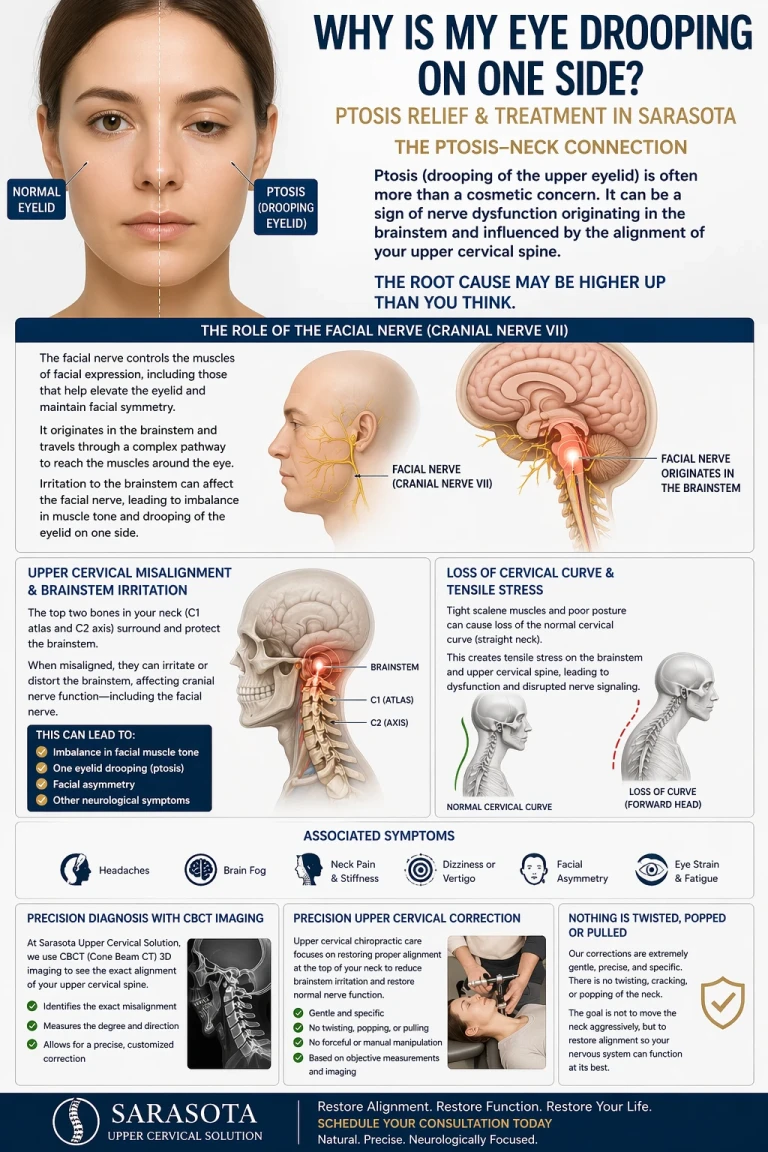
Why Is My Face Drooping on One Side? | Natural Bell’s Palsy Treatment and Relief in Sarasota
Apr 26, 2026
Here is where the mechanism becomes specific, and where the physiology is genuinely well documented — even though the clinical application in chiropractic remains a hypothesis.
Blood pressure regulation on standing is a brainstem operation. Baroreceptors in the carotid sinus and aortic arch detect stretch in the vessel wall and send that information through the glossopharyngeal and vagus nerves into the nucleus tractus solitarius, the NTS, in the dorsolateral medulla. The NTS is the first central relay for cardiovascular afferent information and the integrating hub for the entire baroreflex. From there, excitatory projections run to the caudal ventrolateral medulla, the CVLM, which is inhibitory, and the CVLM in turn restrains the rostral ventrolateral medulla, the RVLM. The RVLM is the principal source of sympathetic drive to the heart and blood vessels, projecting down to the intermediolateral cell column of the spinal cord. In parallel, the NTS drives the parasympathetic side through the nucleus ambiguus and dorsal motor nucleus of the vagus, which slow the heart.
That is the loop. Blood pressure falls on standing, baroreceptor firing drops, NTS output changes, CVLM inhibition of the RVLM is released, sympathetic outflow rises, vessels constrict, heart rate rises modestly, and pressure is maintained. In POTS, that system does not produce a stable outcome — the heart rate rises excessively while pressure is maintained poorly or maintained only at the cost of enormous sympathetic effort.
The relevant question is whether the neck has any input into that circuit. It does.
Bolton, Kerman, Woodring, and Yates demonstrated in 1998 that stimulating the C2 dorsal root ganglion, and the C2 and C3 nerve branches supplying the dorsal neck muscles, produced measurable responses in the splanchnic sympathetic nerve. Neck afferents, in other words, project into circuitry that sets sympathetic outflow. Their work also showed that these cervical influences and vestibular influences on sympathetic output behave antagonistically — arranged so that a whole-body movement produces a cardiovascular response while a head movement on a stationary trunk does not. In human subjects, researchers have since shown that sinusoidal and sustained stretching of neck muscles modulates muscle sympathetic nerve activity directed to the lower limbs, and have concluded that neck proprioceptors may contribute to blood pressure regulation during orthostatic challenge.
Consider what that arrangement is actually for. When you stand up, gravity begins pulling blood toward your legs, and your body must clamp down on the lower-limb vasculature to stop it pooling. When you merely tilt your head back to look at a ceiling, your vestibular system detects head motion in space that looks similar — but no vascular response is needed, because your body has not moved. The brain distinguishes between these two situations by comparing vestibular signals against neck proprioceptive signals. Vestibular input says the head moved. Neck input says whether the body moved with it.
Now imagine that neck input is degraded — noisy, asymmetric, or reporting a position that does not match reality because the joint it is reporting from is not sitting where the brain expects. The comparison becomes unreliable. The system's ability to correctly infer body position from head position is compromised. And the sympathetic response to standing, which depends on that inference, becomes less well calibrated.
That is the mechanistic argument for why an injury that damages upper cervical proprioceptors might contribute to orthostatic intolerance. It does not require the atlas to be pressing on anything. It does not require compression of the brainstem. It requires only that the highest-resolution postural sensor in the body be sending degraded information into a circuit that uses postural information to set cardiovascular tone.
There are secondary considerations layered on top. The vagus nerve exits the skull through the jugular foramen and descends within the carotid sheath immediately anterior to the transverse process of the atlas, which places the primary parasympathetic pathway in close mechanical relationship to the first cervical vertebra. The upper cervical region is also the corridor for venous drainage from the cranium and for cerebrospinal fluid movement at the craniocervical junction, and both of those are relevant to intracranial pressure dynamics, which interact with autonomic regulation. These relationships are anatomically real. How much any of them contributes to a given patient's symptoms is not established, and anyone who tells you otherwise is going further than the evidence goes.
Related article
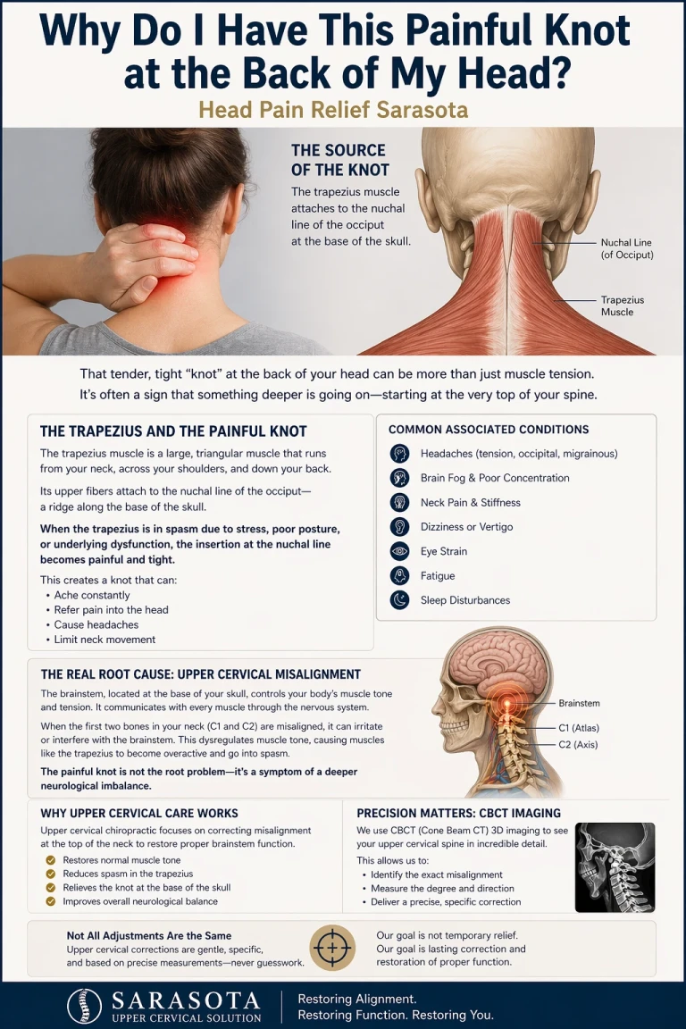
Why do I have this painful knot at the back of my head | Head Pain relief sarasota
Apr 23, 2026What we can and cannot claim
Upper cervical chiropractic research in POTS consists of case reports and small case series. A 2024 case report by Trager and colleagues in Cureus documented a patient with cervicogenic dizziness, upper cervical instability, and POTS whose Dizziness Handicap Inventory score fell from 50 to 10 after eight months of conservative care. A 2022 case study in the Journal of Upper Cervical Chiropractic Research documented resolution of chronic POTS and dysautonomia following upper cervical correction. These are real reports of real patients and they are worth knowing about. They are also not randomized controlled trials, they cannot establish that the intervention caused the improvement, and outcomes documented in a case report should never be presented as what a given person should expect.
It is also true that degenerative and alignment findings in the cervical spine appear frequently in people with no symptoms at all. Finding something on imaging does not by itself establish that the finding explains the illness. Honest evaluation means holding both facts at once: the upper neck is anatomically positioned to influence autonomic regulation, and imaging findings alone do not prove that it is doing so in your case.
What a careful upper cervical evaluation can offer is an assessment of whether the top of your neck is a plausible contributor in your particular situation — using history, orthostatic measurement, examination of upper cervical mechanics and proprioceptive function, and where appropriate three-dimensional cone beam CT imaging to assess the actual alignment relationship between the skull, atlas, and axis. In post-traumatic cases specifically, the questions that raise suspicion are straightforward. Did symptoms begin after the injury rather than before it? Is there persistent upper neck pain, suboccipital tightness, or headache alongside the autonomic symptoms? Is there dizziness that changes with head position? Has anyone actually examined the upper cervical spine, as opposed to imaging the brain and declaring it normal?
That last question is the one that most often has not been asked. After a concussion, attention goes to the brain. After a car accident, attention goes to fractures and disc pathology. The craniocervical junction — the region that took the shear load, that houses the densest proprioceptive field in the body, that sits directly beneath the brainstem nuclei running the baroreflex — routinely goes unexamined by anyone.
If your POTS started after a crash, a fall, or a hit, that gap is worth closing. Not because the neck is guaranteed to be the answer, but because it is a genuinely plausible contributor that is straightforward to evaluate and that almost nobody has looked at.
Upper cervical care is a complementary approach. It is not a replacement for your cardiologist, neurologist, or autonomic specialist, and nothing here is a reason to stop treatment that is working for you.
If you developed POTS or orthostatic intolerance after a car accident, concussion, or neck injury and want to find out whether your upper cervical spine is part of the picture, schedule a free consultation or call 941-259-1891.
Medical disclaimer: This article is for educational purposes only and does not constitute medical advice, diagnosis, or treatment. POTS is a complex condition requiring evaluation by qualified medical professionals. Upper cervical chiropractic care is a complementary approach and is not a substitute for medical management. Individual results vary, and the outcomes described in published case reports should not be interpreted as typical or expected. Always consult your physician before making changes to your care.
Dr. Drew Ahall, Upper Cervical Chiropractor [www.neckwise.com](https://www.neckwise.com)


Leave a comment