
When patients hear the words “upper cervical chiropractic,” they often think about the adjustment itself. But in precise upper cervical care, the adjustment is only one part of the process.
Before care begins, the doctor needs to understand the position, structure, and mechanics of the upper neck in detail.
Schedule Your Appointment
Schedule appointmentThat is where imaging and analysis become important.
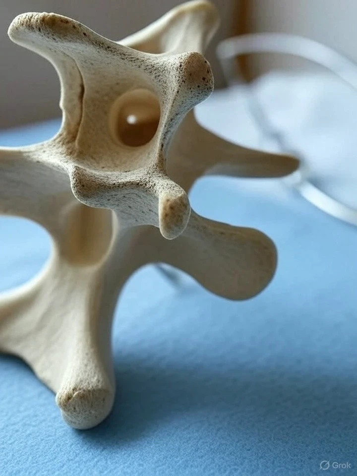
Upper cervical chiropractic focuses on the relationship between the head, the atlas, the axis, and the rest of the spine. The atlas is the first cervical vertebra, also called C1, and the axis is the second cervical vertebra, also called C2.
These two bones sit directly beneath the skull and play a major role in head balance, neck motion, posture, and spinal function.
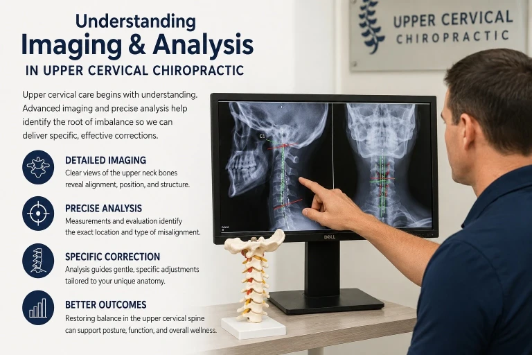
Because this area is small, complex, and highly individual from one person to another, upper cervical chiropractors often use detailed imaging and biomechanical analysis to guide care.
The goal is not to guess where the correction should be made. The goal is to evaluate the patient carefully, measure what can be measured, and deliver care as specifically as possible.
Why Imaging Matters in Upper Cervical Chiropractic
The upper cervical spine is not like the lower back or mid-back. It has a unique shape and function. The atlas does not have a typical vertebral body like many other spinal bones. Instead, it acts more like a ring that supports the head and allows movement between the skull and neck.
Because of this anatomy, small changes in alignment or motion may influence how the head rests over the spine. This can affect posture, muscle tension, joint stress, and movement patterns throughout the body.
Imaging allows the doctor to see structural details that cannot be fully understood by touch alone. A physical examination, posture evaluation, range-of-motion testing, neurological screening, and symptom history are all important.
But imaging can provide an added layer of information about the bony structure of the upper neck.
In upper cervical chiropractic, imaging may help evaluate:
- The position of the atlas and axis
- The relationship between the skull and upper cervical spine
- Head tilt or rotation patterns
- Structural asymmetry
- Joint positioning
- Degenerative changes
- Prior injury patterns
- Anatomical variations
A review of upper cervical chiropractic procedures notes that different upper cervical techniques use different analysis methods, but they commonly examine the relationship between the occiput, atlas, and axis when indicated by evaluation findings.
Imaging Is Not Just About “Seeing the Neck”
One of the most important things patients should understand is that imaging in upper cervical chiropractic is not simply about taking a picture. The image must be taken correctly, positioned accurately, and analyzed with a specific purpose.
Poor positioning can affect measurements. If the patient’s head, body, or equipment is not aligned properly during the image, the final view may create distortion. This is why upper cervical imaging protocols often place strong emphasis on patient positioning and repeatability.
Related article

Upper cervical radiographic education sources explain that precise equipment alignment and proper patient placement are essential for distortion-controlled images that can be accurately analyzed.
For the patient, this means the imaging process may feel more detailed than a standard quick X-ray. The doctor or imaging technician may take time to position the head, shoulders, and neck carefully. This attention to detail is part of the analysis process.
What the Doctor Is Looking For
Upper cervical imaging is used to help answer several important clinical questions.
The doctor may be looking at whether the atlas appears shifted, rotated, tilted, or positioned in a way that may affect the balance of the head and neck. The doctor may also evaluate how the upper cervical spine relates to the lower cervical spine and overall posture.
This does not mean every symptom is caused by one bone being “out of place.” The body is more complex than that. Pain, headaches, dizziness, neck stiffness, postural strain, and nerve irritation can have many causes. Upper cervical analysis is one part of a complete clinical picture.
The imaging findings should be considered alongside:
- The patient’s health history
- Current symptoms
- Previous injuries or accidents
- Neurological findings
- Muscle tone and posture
- Range of motion
- Functional movement patterns
- Response to previous care
This is why imaging alone does not create a treatment plan. It supports the doctor’s clinical reasoning.
X-Rays, CBCT, and Upper Cervical Analysis
Traditional X-rays have long been used in chiropractic and upper cervical analysis. They provide two-dimensional views of the spine and can help evaluate bony alignment, posture, and structural changes.
Some upper cervical offices may also use cone beam computed tomography, commonly called CBCT. CBCT provides three-dimensional imaging and may offer more detailed visualization of the craniovertebral junction, the area where the skull and upper cervical spine meet.
Research and clinical discussion have noted that CBCT can provide improved visualization of certain craniocervical structures compared with traditional radiography in some contexts.
The type of imaging used depends on the office, the doctor’s technique, the patient’s case, and whether imaging is clinically appropriate.
Not every patient needs the same imaging. A responsible chiropractor considers the patient’s age, history, symptoms, exam findings, prior imaging, radiation exposure, and clinical need before recommending imaging.
The Role of Measurement
Upper cervical chiropractic is often described as precise because many techniques use measurements to guide the correction. Instead of applying a general adjustment to the neck, the doctor uses imaging analysis to determine direction, angle, and correction strategy.
Related article
Upper Cervical Chiropractic: How Atlas Adjustments Can Transform Your Life
Jun 01, 2025This may include evaluating:
- Atlas laterality
- Atlas rotation
- Head tilt
- Cervical curve changes
- Occiput-atlas-axis relationship
- Structural asymmetry
- Pre- and post-correction comparison
The purpose of measurement is to make care more specific to the individual patient. Two patients may both have neck pain, but their upper cervical findings may be very different. One patient may show more rotation. Another may show more head tilt. Another may have a history of trauma that affects how the spine compensates.
That is why upper cervical care is not usually a one-size-fits-all approach.
Why Post-Analysis Matters
In some upper cervical techniques, follow-up analysis may be used to evaluate whether the correction changed the measured alignment or improved the mechanical relationship of the upper neck.
This does not mean the goal is to constantly adjust the patient. In fact, many upper cervical approaches focus on helping the correction hold for as long as possible. The idea is not to adjust more often than needed. The idea is to monitor the patient’s progress and determine whether the body is maintaining better balance.
This may include checking posture, leg balance, muscle tone, range of motion, neurological indicators, and symptom changes.
When imaging is used before and after care, it may help the doctor compare structural findings and determine whether the correction had the intended mechanical effect.
Imaging and Safety
Patients sometimes ask whether imaging is safe. This is an important question.
Any form of X-ray or CT-based imaging involves radiation exposure. That is why imaging should be recommended only when there is a clinical reason. The benefit of the information should outweigh the exposure involved.
Doctors who use imaging should follow appropriate radiographic safety principles, including proper positioning, shielding when applicable, and avoiding unnecessary repeat imaging. Chiropractic radiography discussions emphasize that imaging should be part of clinical decision-making rather than used casually or without purpose.
For patients, the best approach is to ask questions:
- Why is this imaging recommended?
- What information are you looking for?
- How will the findings affect my care plan?
- Have my prior images been reviewed?
- Are there alternatives if imaging is not appropriate for me?
A good clinical conversation should help the patient understand the reason behind the recommendation.
What Patients Can Expect During Upper Cervical Imaging
While every office may have a different process, many patients can expect a careful setup. The doctor or staff may position the head and neck in specific ways to capture the needed views.

The process is usually not painful, though patients with acute neck pain may feel some stiffness while holding still.
The imaging process may include views that focus on the upper cervical area rather than only standard neck views. These views are intended to show the relationship between the skull, atlas, axis, and surrounding structures.
After the images are taken, the doctor performs an analysis. This may involve measurements, line drawings, digital software, or technique-specific protocols.
The findings are then used to help determine whether upper cervical care is appropriate and how the correction should be delivered.
Why This Matters for Patients in Sarasota
For patients searching for upper cervical chiropractic care in Sarasota, imaging and analysis can help explain why this type of care is different from a general neck adjustment. The focus is on understanding the individual structure of the upper neck before applying a correction.
Patients often seek upper cervical care for concerns such as neck pain, headaches, postural imbalance, dizziness, tension, and symptoms that may relate to prior injuries or long-term spinal stress.
Imaging does not diagnose every cause of these symptoms, but it may help the doctor better understand the structural and mechanical factors involved.
At Sarasota Upper Cervical, this topic matters because patients deserve a thoughtful, measured, and individualized approach. The upper neck is a delicate and important region. Care should be based on evaluation, not assumption.
Common Questions About Upper Cervical Imaging
Is imaging always required before upper cervical chiropractic care?
Not always. The need for imaging depends on the doctor’s clinical judgment, the patient’s history, examination findings, and the technique used. Some cases may require imaging for safety and precision, while others may not.
Can imaging show nerve pressure?
Standard X-rays show bones and alignment, not nerves directly. Advanced imaging such as MRI is usually better for evaluating discs, nerves, and soft tissues. Upper cervical imaging is generally used to analyze structure, alignment, and joint relationships.
Why does positioning matter so much?
Small errors in positioning can affect how the image appears. Since upper cervical analysis may involve precise measurements, accurate positioning helps reduce distortion and improves the reliability of the analysis.
Does an upper cervical adjustment hurt?
Upper cervical corrections are often gentle and specific. Many techniques use low-force methods rather than twisting or forceful manipulation. The exact experience depends on the technique and the patient’s condition.
What happens after the analysis?
The doctor reviews the findings and explains whether upper cervical care may be appropriate. If care begins, the analysis helps guide the direction and type of correction.
Final Thoughts
Imaging and analysis are central parts of many upper cervical chiropractic approaches because they help the doctor understand the patient’s individual anatomy and spinal mechanics. The goal is precision, not guesswork.
For patients, this means care should begin with a complete evaluation. Imaging, when clinically appropriate, can provide important information about the upper neck and help guide a more specific correction.
If you are dealing with neck pain, headaches, posture problems, or symptoms that may be connected to upper cervical stress, a detailed evaluation may help determine whether upper cervical chiropractic care is appropriate for you.
To learn more or schedule a consultation, visit Sarasota Upper Cervical at sarasotauppercervical.com.


Leave a comment