

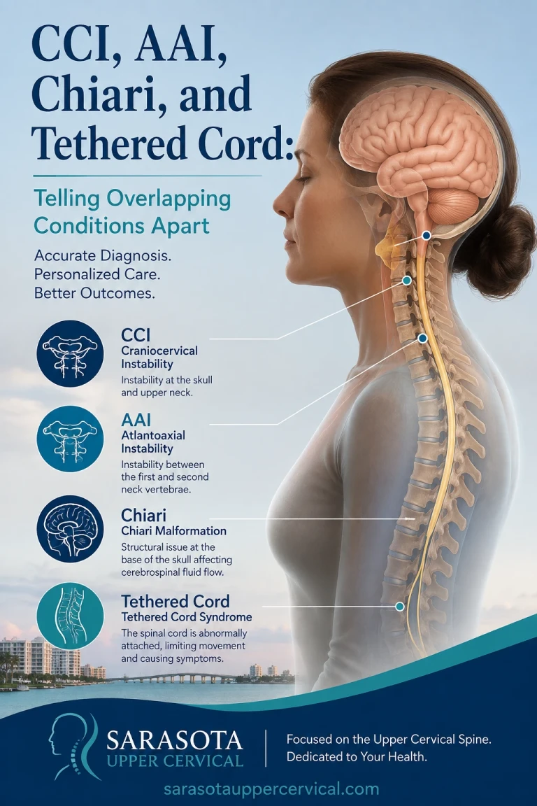
CCI, AAI, Chiari, and Tethered Cord: Telling Overlapping Conditions Apart
Four conditions come up repeatedly in the same conversations, in the same patient communities, and often in the same patients. They get used almost interchangeably online, which is understandable — the symptoms overlap heavily and they genuinely do co-occur — but they are distinct problems in distinct locations with distinct treatments.
Schedule Your Appointment
Schedule appointmentGetting them confused has consequences. People pursue evaluation for the wrong one, get a negative result, and conclude nothing structural is going on. Or they attribute everything to whichever name they encountered first and stop looking. This article separates them.
A caution before starting: all four are medical diagnoses. None of them are diagnosed by a chiropractor, including at this practice. What follows is intended to help you understand your own situation well enough to ask better questions of the physicians who do diagnose them.
Craniocervical instability
Where: Between the skull and the atlas, and between the atlas and the axis — the occiput-C1 and C1-C2 levels.
What: Excess motion resulting from insufficient ligamentous restraint. The craniocervical junction has minimal bony stability by design, trading it for range of motion, and relies on the transverse ligament, the alar ligaments, and the tectorial membrane to limit movement. When those tissues are lax or damaged, the skull moves relative to the upper cervical spine beyond physiological range.
Why it causes symptoms: Not the motion itself, but what the motion does to structures passing through a crowded space — the brainstem and upper spinal cord, the vertebral arteries, the jugular veins, cranial nerves nine through twelve, and cerebrospinal fluid pathways. Deformative stress on the brainstem and upper cord is the mechanism of greatest concern.
Typical picture: Positional headache at the skull base, worse upright and better lying flat. A heavy head sensation. Cranial nerve symptoms clustering together — swallowing difficulty, tinnitus, voice changes, visual disturbance. Autonomic symptoms. Cognitive fog that tracks with upright time.
How it is identified: Upright dynamic imaging in flexion, extension and neutral, with craniometric measurement. Lohkamp, Marathe and Fehlings, in their 2022 systematic review in Global Spine Journal, recommended four parameters: the clivo-axial angle, the Harris measurement, the Grabb-Mapstone-Oakes measurement, and the angular displacement of C1 on C2. The clivo-axial angle measures how sharply the brainstem must bend passing from skull into neck, with normal values around 155 degrees and angles at or below roughly 135 degrees considered potentially pathological. The Grabb-Mapstone-Oakes measurement assesses ventral encroachment on the canal.
Atlantoaxial instability

Where: Specifically at C1-C2.
What: Excess motion at the atlantoaxial joint — most often anterior translation of the atlas on the axis when the transverse ligament is insufficient, or excess rotation when the alar ligaments are. AAI can exist alone or as part of a broader craniocervical instability picture, and the terms are often used loosely, but the anatomical distinction is real: CCI is a broader problem including the skull-atlas relationship, AAI is specific to the atlas-axis joint below it.
Why it matters separately: The C1-C2 joint accounts for roughly half of all cervical rotation, and the spinal cord passes directly through the ring of C1. It also has causes that CCI in the connective-tissue sense does not — rheumatoid arthritis erodes the transverse ligament and the odontoid over time, Down syndrome is associated with congenital laxity at this level, and trauma can rupture the transverse ligament outright.
Typical picture: Substantially overlaps with CCI, with rotational symptoms often more prominent — dizziness or symptom provocation on turning the head, upper neck pain localized to one side, occipital headache.
Evidence worth knowing: Henderson, Rowe, Narayanan and colleagues published a study in World Neurosurgery in 2021 on refractory syncope and presyncope associated with atlantoaxial instability, describing improvement following surgical stabilization in patients whose fainting had persisted despite maximal medical management. That study is the strongest single piece of evidence linking this specific joint to autonomic dysfunction.
How it is identified: Flexion-extension imaging measuring the atlanto-dental interval — the gap between the odontoid and the anterior arch of C1 — with reported upper limits in the literature around 3 mm in men and 2.5 mm in women. Rotational CT is used for rotational instability.
Chiari malformation type I
Where: At the foramen magnum, but the problem is in the skull rather than the joints.
What: The cerebellar tonsils descend below the level of the foramen magnum into the upper spinal canal. This is a structural anomaly of the posterior fossa — often the posterior fossa is simply too small for its contents — rather than a problem of joint motion.
Why it causes symptoms: Crowding at the foramen magnum obstructs cerebrospinal fluid flow between the cranial and spinal compartments and can compress the brainstem and cerebellum directly. In some cases a syrinx — a fluid cavity within the spinal cord — develops as a consequence.
Typical picture: The classic feature is a headache provoked by Valsalva maneuvers — coughing, sneezing, straining, laughing — which is more specific to Chiari than to the instabilities. Otherwise it overlaps heavily: occipital headache, dizziness, swallowing difficulty, visual disturbance, tinnitus, and sensory changes.
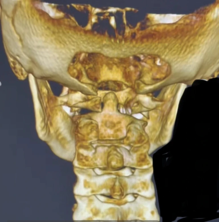
The critical overlap: Chiari and craniocervical instability frequently coexist in connective tissue disorders, and this is well described. Milhorat and colleagues characterized a syndrome of occipitoatlantoaxial hypermobility, cranial settling, and Chiari type I malformation in patients with hereditary disorders of connective tissue in the Journal of Neurosurgery: Spine in 2007. Cranial settling — the skull descending on the cervical spine as ligaments become insufficient — can produce or worsen tonsillar descent, which means the two conditions are mechanically related rather than merely co-occurring. This matters surgically, because decompressing a Chiari without addressing underlying instability can worsen the instability.
How it is identified: Standard brain MRI, measuring tonsillar descent below the foramen magnum. Notably, this is the one condition on this list that a routine supine MRI does detect reasonably well.
Tethered cord syndrome
Where: The opposite end of the spine — the filum terminale at the base of the spinal cord.
What: The spinal cord is abnormally anchored at its lower end, restricting its normal movement within the canal and placing traction on the cord.
Why it belongs in this conversation: Because it co-occurs with cervical instability in hypermobile EDS, and because that co-occurrence is under-recognized. Gensemer, Daylor, Nix, Norris and Patel addressed exactly this in their 2024 review in Frontiers in Neurology, noting that hEDS patients may experience spinal neurological complications including cervico-medullary symptoms arising from cranio-cervical and cervical instability, as well as tethered cord syndrome — and emphasizing that TCS is often radiographically occult in nature, not always detectable on standard imaging.
Typical picture: Distinct from the others and located lower — lower back pain, balance difficulty, weakness in the legs, sensory loss, and bowel or bladder dysfunction. If your symptom picture includes leg weakness and bladder changes alongside the upper cervical complaints, this is the condition that accounts for the lower half.
How it is identified: Difficult, precisely because the occult form is not reliably visible on imaging. Diagnosis often rests substantially on clinical presentation, and MRI of the whole spine is recommended primarily to exclude other causes of back pain and leg weakness. Surgical management involves sectioning the filum terminale, and re-tethering after surgery is possible.
How to tell them apart in practice
Location of symptoms is the first sort. Predominantly head, neck, cranial nerve and autonomic symptoms point upward, toward CCI, AAI, or Chiari. Lower back, leg weakness, sensory loss and bladder or bowel changes point downward, toward tethered cord. Both together suggests both, which in hEDS is common.
Positional pattern helps sort the upper three. Symptoms clearly worse upright and better lying flat point toward instability or toward a cerebrospinal fluid leak. Symptoms provoked specifically by coughing, sneezing or straining point toward Chiari. Symptoms provoked by head rotation specifically point toward the atlantoaxial joint.
Related article
Chiari Malformation Healing After Surgery: A Sarasota Upper Cervical Success Story
Apr 14, 2026Imaging modality determines what can even be found. Chiari shows on a standard supine MRI. Instability generally does not — it requires upright dynamic imaging with craniometric measurement. Occult tethered cord may not show at all. A normal brain MRI meaningfully reduces the likelihood of Chiari; it says very little about the other three.
Assume overlap rather than exclusivity. This is the most useful single principle. In hypermobile connective tissue disorders these conditions travel together, and finding one is a reason to evaluate for the others rather than a reason to stop looking. A great deal of diagnostic delay in this population comes from stopping at the first finding.
Being honest about the state of the evidence
This is contested territory within medicine and you should know that going in. Lohkamp and colleagues found only sixteen qualifying articles covering seventy-eight surgical patients in their systematic review, concluding there is a lack of high-quality prospective evidence for evaluating suspected CCI in EDS, and recommending that surgical fixation be performed only where there is clear radiographic instability together with concordant symptoms and signs. Mao and colleagues published a 2022 paper in The Spine Journal organized entirely around the controversies. Diagnostic thresholds, imaging protocols and surgical indications are all subjects of genuine disagreement among the specialists who work in this area.
That does not mean these conditions are not real. It means the field is young, the criteria are still moving, and both over-diagnosis and under-diagnosis occur. A physician who is cautious here may be reading the same literature you are.
Where an upper cervical practice fits
Narrowly, and it is worth being explicit about the boundary.
We evaluate upper cervical alignment and mechanics, including three-dimensional cone beam CT imaging of the alignment relationship between skull, atlas and axis. That assesses alignment. It does not diagnose craniocervical instability, atlantoaxial instability, Chiari malformation, or tethered cord syndrome — all four of which are medical diagnoses requiring physician evaluation and, in three cases, imaging we do not perform.
What an evaluation here contributes is a structural assessment of the upper cervical spine, identification of mechanical dysfunction that may be contributing to your symptoms, and — importantly — a judgment about whether hands-on care is appropriate for you at all. In frank instability, high-velocity cervical manipulation is contraindicated, and in someone with a suspected structural diagnosis the correct first step is medical referral rather than treatment.
If you are trying to work out which of these four you might be dealing with, the most useful thing is usually to get clear on what has actually been imaged and measured so far. That is frequently less than people assume.
If you are trying to sort out overlapping upper cervical symptoms and want a structural evaluation along with a clear assessment of whether medical referral is your right next step, schedule a free consultation or call 941-259-1891.
Medical disclaimer: This article is for educational purposes only and does not constitute medical advice, diagnosis, or treatment. Craniocervical instability, atlantoaxial instability, Chiari malformation, and tethered cord syndrome are all medical diagnoses requiring evaluation by qualified physicians using appropriate imaging. Cervical manipulation may be contraindicated in the presence of instability. Progressive neurological symptoms require prompt medical evaluation. Upper cervical chiropractic care is a complementary approach and is not a substitute for medical or surgical management. Individual results vary. Always consult your physician before making changes to your care.


Leave a comment