

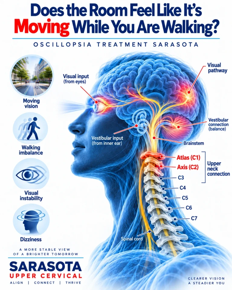
Oscillopsia is the sensation that the visual world is moving when it is not, and people in Sarasota, Bradenton, and Lakewood Ranch who experience it usually struggle to describe it. The room bounces when walking. Signs jitter. Faces will not settle. Reading is impossible while moving. It is a distinct symptom from blurred vision and from dizziness, and identifying which of several very different conditions is producing it determines everything that follows.
This article is largely about that differential, because oscillopsia has causes ranging from benign to serious, and one of the least likely explanations is the neck.
Schedule Your Appointment
Schedule appointmentWhy the world normally stays still
Every time you take a step, your head moves. The image on your retina moves with it. That you do not perceive the world as bouncing is the achievement of the vestibulo-ocular reflex, which detects head movement through the semicircular canals and drives compensatory eye movement in the opposite direction at matched velocity, with a latency of only a few milliseconds — far faster than any visually guided correction could manage.
Oscillopsia occurs when that stabilisation fails. Either the reflex is not generating adequate compensation, or the eyes are moving when they should be still.
The main causes, and how they differ
Bilateral vestibular loss
When both inner ears lose function, the vestibulo-ocular reflex cannot compensate for head movement. The characteristic pattern is oscillopsia present only during movement — walking, running, riding in a car — with vision stable when still. Patients often report they must stop walking to read a sign. Balance is markedly worse in the dark, when vision can no longer substitute for absent vestibular input.
Aminoglycoside antibiotics, particularly gentamicin, are a well-recognised cause, as are bilateral vestibular neuritis, Ménière's disease affecting both ears, and some genetic and autoimmune conditions. This diagnosis is frequently delayed by years, and it is worth asking directly about a history of intravenous antibiotic treatment.
Nystagmus
Where the eyes drift and correct involuntarily, the image moves across the retina and produces oscillopsia at rest as well as during movement. Acquired nystagmus in an adult always warrants neurological evaluation, because the causes include multiple sclerosis, stroke, tumour, medication effects, and Chiari malformation. Downbeat nystagmus in particular points toward the craniocervical junction and cerebellum, and requires imaging.
Congenital nystagmus is different — people who have had it since infancy usually do not experience oscillopsia, because the visual system adapted early.
Superior canal dehiscence
A third window in the bone over the superior semicircular canal produces oscillopsia triggered by loud sound or by pressure change from coughing, sneezing, or straining, with vertical-torsional eye movement aligned to the plane of that canal. It also produces distinctive auditory features — autophony, and hearing internal body sounds such as one's own eye movements or footsteps. Pulsatile oscillopsia, the visual field bouncing with the heartbeat, appears here too.
Unilateral vestibular loss
Loss on one side produces oscillopsia mainly on rapid head turns toward the affected side, and it usually improves as central compensation develops.
Ocular and other causes
Superior oblique myokymia produces brief episodes of monocular shimmering. Some medications, including certain anticonvulsants and lithium, cause it. Thyroid eye disease and orbital problems can produce unstable vision through different mechanisms.
Where the cervical spine fits — and where it does not
I want to be direct, because oscillopsia is a symptom that attracts confident cervical explanations and most of them are wrong.
Oscillopsia in its classic forms is a vestibular or ocular motor problem. Bilateral vestibular loss, nystagmus, and superior canal dehiscence are not neck conditions, and no amount of upper cervical care addresses them. Someone with progressive oscillopsia needs vestibular function testing and, where indicated, neuroimaging — not a course of adjustments.
What is legitimate is narrower. Cervical afferents from C1 to C3 project into the vestibular nuclei and contribute to the cervico-ocular reflex, which works alongside the vestibulo-ocular reflex in gaze stabilisation. Where vestibular function is reduced, the cervico-ocular reflex has been described as contributing more, and the quality of cervical proprioceptive input becomes correspondingly more relevant. This is why sensorimotor retraining forms part of vestibular rehabilitation.
Cervical injury can also produce visual instability that patients describe in oscillopsia-like terms. The smooth pursuit neck torsion test, studied by Julia Treleaven and colleagues at the University of Queensland, demonstrated significantly disturbed eye tracking when the trunk was rotated beneath a stationary head in people with persistent whiplash compared with controls, with the abnormality present even in patients who did not report dizziness. Findings across research groups have not been uniform, and the test is a useful measure rather than a settled standard.
The honest position is that a cervical contribution to gaze instability exists and is measurable, that it is not the same thing as true oscillopsia from vestibular failure, and that establishing which one a patient has must come before any treatment decision. There is no trial of upper cervical chiropractic for oscillopsia.
Related article
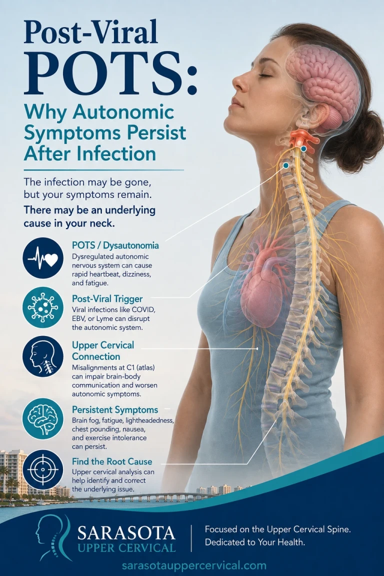
Post-Viral POTS: Why Autonomic Symptoms Persist After Infection | Sarasota
Aug 02, 2026How the distinction is made
The history does most of the work. Oscillopsia only during head movement, with stable vision at rest, points toward vestibular loss. Oscillopsia at rest points toward nystagmus. Oscillopsia triggered by loud sound or straining points toward a third window. Oscillopsia that varies with neck position and comes with neck pain and restricted rotation raises the cervical possibility — but it should be a diagnosis of exclusion here, not a first assumption.
Formal assessment involves vestibular function testing, video head impulse testing, dynamic visual acuity testing, examination for nystagmus in various gaze positions, and imaging where indicated. Those belong with a neurologist, neurotologist, or vestibular specialist.
Precision and knowing when not to adjust
Where cervical involvement is genuinely identified, correction involves no twisting, no popping, no cracking, and no pulling of the neck. Cone beam computed tomography measures the three-dimensional position of the atlas and axis against your own anatomy so a correction is calculated rather than estimated. Objective measures — leg length assessment, cervical range of motion, joint position error testing, balance measurement including tandem stance with eyes closed, and thermographic pattern tracking — determine whether a correction is indicated and whether it is holding. When it is holding, no adjustment is given.
Cone beam imaging of the cervical spine does not assess vestibular function and should never be presented as though it does. Where the history suggests a vestibular or central cause, the appropriate outcome of an evaluation here is a referral.
An evaluation in Sarasota
If the world moves when you do, the first priority is establishing why, which usually means vestibular testing and neurological assessment. Where those have been completed and a cervical contribution to gaze instability remains a live question — particularly after whiplash or head injury — an upper cervical evaluation with sensorimotor testing can assess it. Where the picture suggests a vestibular or central cause, you will leave with a referral rather than a treatment plan. To arrange a consultation, visit NeckWise.
Dr. Drew Hall, Upper Cervical Chiropractor
Serving Sarasota, Bradenton, and Lakewood Ranch, Florida
This article is for general educational purposes only and is not medical advice, a diagnosis, or a treatment recommendation for any individual. Oscillopsia requires evaluation by qualified medical providers, typically including vestibular function testing and neurological assessment, and acquired nystagmus in an adult requires neuroimaging. Upper cervical chiropractic is an area of focus within chiropractic; it is not a board-recognized specialty, and no claim of specialization or superiority is made or implied. Upper cervical care does not treat bilateral vestibular loss, nystagmus, or superior canal dehiscence, and no such claim is made here; no trial evidence exists for upper cervical chiropractic in oscillopsia, and findings on the smooth pursuit neck torsion test have not been uniform across research groups. Cone beam computed tomography is used as a precision measurement and analysis tool, not as a stand-alone diagnostic device, and does not assess vestibular function. Individual results vary and no specific outcome is guaranteed. Seek emergency care for sudden oscillopsia with headache, weakness, numbness, slurred speech, facial droop, or difficulty swallowing. Always consult a qualified healthcare provider regarding diagnosis and treatment.


Leave a comment