

Introduction
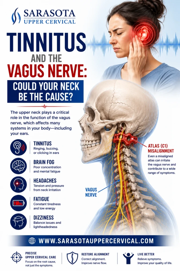
Tinnitus, a persistent ringing, buzzing, whooshing or hissing sound with no external source — is usually attributed to hearing damage, and in many cases that's accurate. But a well-documented subset of tinnitus, sometimes called somatic tinnitus or cervicogenic tinnitus, has nothing to do with the ear at all. It originates from the neck, and the anatomical bridge between the two is the same structure at the center of nearly every vagus nerve discussion: the tight cluster of nerves, vessels, and joints packed into the upper cervical spine.
The Anatomy: Where Hearing, the Vagus Nerve, and the Upper Neck Overlap
Schedule Your Appointment
Schedule appointment
The reason neck dysfunction can produce a sound with no auditory cause comes down to a shared brainstem structure: the dorsal cochlear nucleus, the first relay station for auditory signals coming from the inner ear. This nucleus doesn't only receive input from the ear, it also receives direct convergent input from the upper cervical spinal cord (particularly the dorsal column and spinal trigeminal nucleus) and, notably, from vagal and other cranial nerve afferents that pass through the same brainstem region.
This convergence means the auditory processing centers of the brainstem are not isolated from neck and vagal signaling — they are wired into the same circuitry. When the upper cervical spine sends abnormal or excessive sensory input into the brainstem (from joint dysfunction, muscle tension, or nerve irritation), that input can spill over into auditory processing centers and be perceived as sound, even though nothing is wrong with the ear itself.
The vagus nerve is directly relevant here for two anatomical reasons:

Its exit point. The vagus nerve exits the skull through the jugular foramen, immediately adjacent to the internal auditory canal and the temporal bone structures housing the inner ear. The glossopharyngeal nerve (which also contributes sensory input to the ear via the tympanic branch) exits through the same foramen. Mechanical crowding or tension at this exit point — from upper cervical misalignment pulling on the surrounding fascia — can affect multiple cranial nerves simultaneously, not just one in isolation.
Its course through the neck. After exiting the skull, the vagus nerve travels within the carotid sheath directly past the atlas (C1) and axis (C2), alongside the internal jugular vein. The middle ear and inner ear structures share venous drainage pathways with this same jugular system. Restriction of jugular venous outflow — which can occur with upper cervical misalignment affecting the fascia around the jugular foramen — has been associated with pressure changes that some researchers believe contribute to pulsatile and non-pulsatile tinnitus.
Why C1 and C2 Are the Key Structures
The atlas and axis are the only two vertebrae in the body that directly articulate with the skull, and they do so through joints with an extremely high density of proprioceptive nerve endings, far more concentrated than any other spinal region. These joints feed directly into the cervical spinal nucleus, which converges with the cochlear nucleus in the brainstem.
When C1 or C2 is misaligned, a common and frequently undiagnosed consequence of whiplash, a fall, sports injury, or long-term postural strain, several things can happen at once:
Abnormal proprioceptive signaling
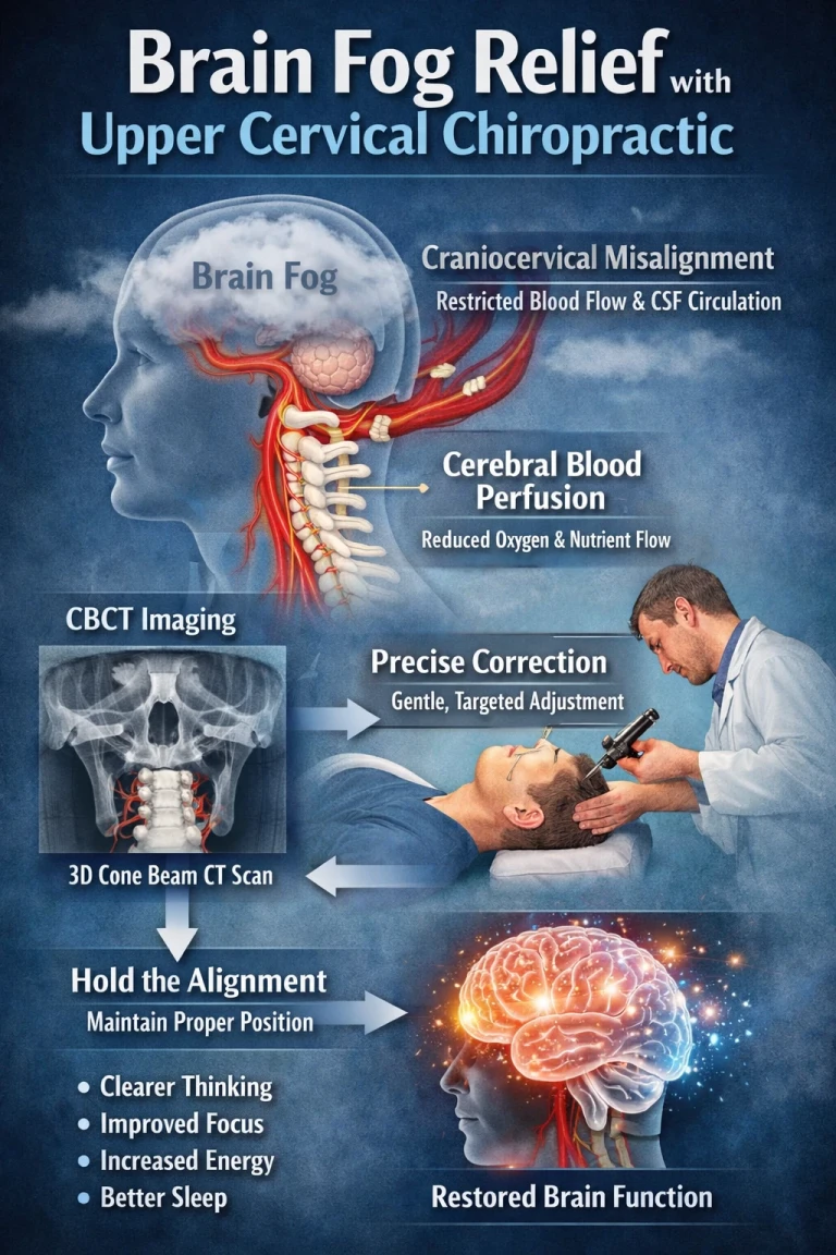
floods into the cervical spinal nucleus and spills into the adjacent cochlear nucleus, potentially generating phantom auditory signals
Fascial tension along the carotid sheath and jugular foramen may mechanically affect the vagus nerve and the venous drainage it runs alongside, altering local pressure dynamics near the inner ear's venous outflow
Suboccipital muscle tension, which is common with upper cervical misalignment, can refer pain and altered sensory signaling into the same brainstem region processing both neck position and auditory input
Reduced vagal tone overall may lower the nervous system's capacity to filter and dampen aberrant sensory signals — vagal tone plays a broader role in sensory gating, which is the brain's ability to suppress irrelevant or non-threatening background signals (a mechanism thought to be impaired in many tinnitus cases regardless of origin)
This is why cervicogenic tinnitus often correlates with, and sometimes fluctuates in intensity alongside, neck stiffness, headaches, or a history of whiplash, and why it frequently does not respond to standard audiological treatment, since the auditory system itself may not be the source of the problem.
Vagal Tone and Sensory Gating
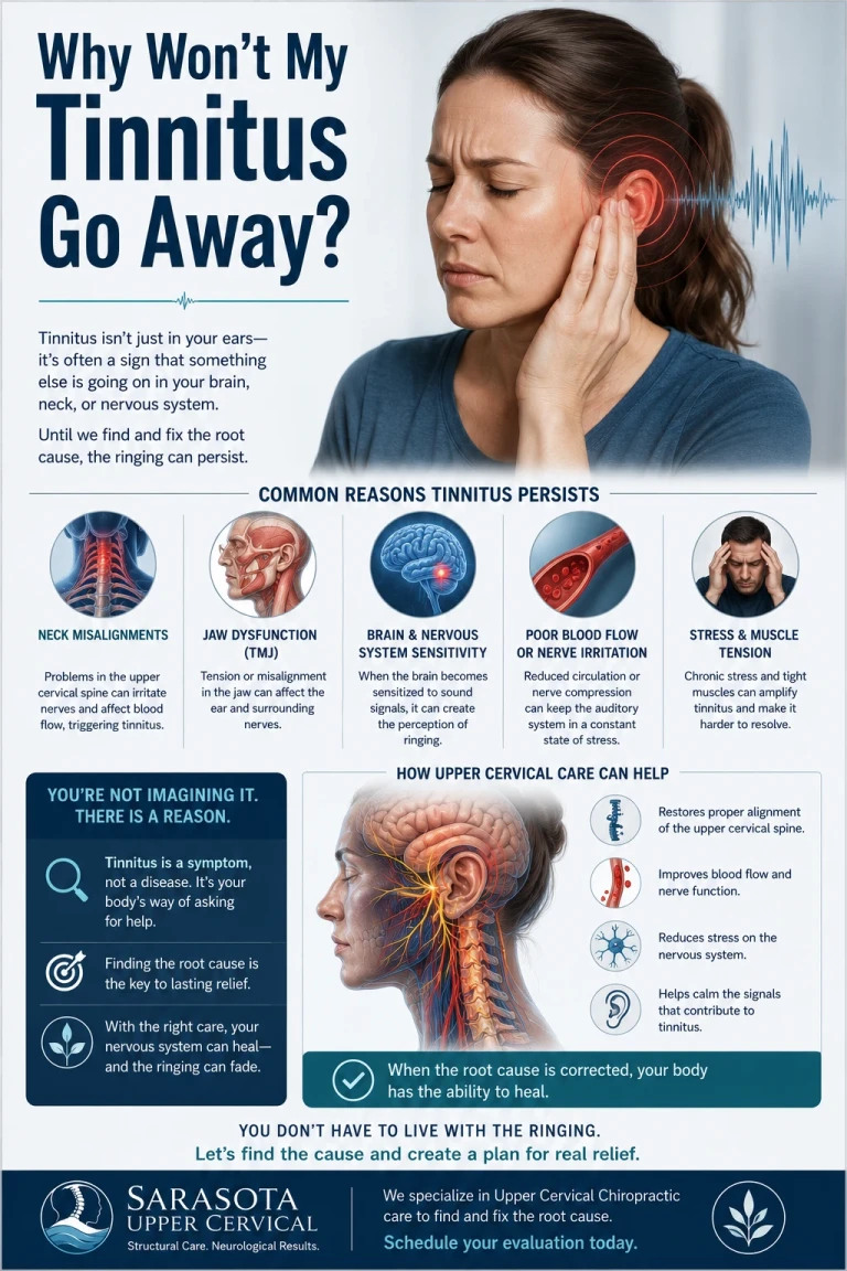
Beyond the direct anatomical proximity, the vagus nerve plays a broader regulatory role relevant to tinnitus: it's a central player in autonomic and sensory gating, the nervous system's capacity to distinguish signal from noise and suppress background input that isn't relevant. Reduced vagal tone has been associated with:
1.Heightened overall nervous system arousal, which can amplify the perceived loudness and distress of tinnitus even if the underlying phantom signal itself doesn't change
2.Poor stress recovery, and stress is one of the most consistently reported factors that worsens tinnitus intensity
3.Disrupted brainstem-level filtering of low-level sensory noise, which is part of the leading theoretical model for why tinnitus becomes consciously perceived and persistent in some people rather than fading into the background the way most background neural noise does
What This Means in Practice
Standard tinnitus care — hearing evaluation, sound therapy, hearing aids where indicated — remains essential when there is an underlying auditory or hearing-loss component. But when tinnitus doesn't correlate with measurable hearing loss, or fluctuates with neck position, stress, or a history of head or neck trauma, the upper cervical spine and its relationship to the vagus nerve and brainstem auditory pathways deserves consideration as part of a complete picture.
Upper cervical specific chiropractic care uses precision imaging to identify the exact position of C1 and C2 relative to the skull and spine, and delivers a specific, measured correction aimed at removing mechanical interference at the exact junction where the cervical spinal nucleus, the vagus nerve, and the brainstem's auditory processing centers converge.
If your tinnitus is accompanied by neck stiffness, headaches, a history of whiplash, or fluctuates with head and neck position, it may be worth having your upper cervical alignment evaluated.


Leave a comment