

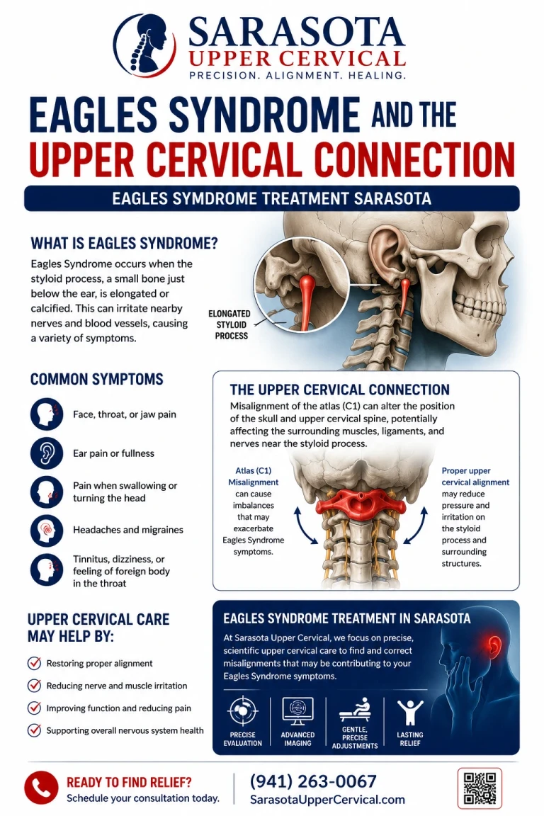
Eagle syndrome treatment in Sarasota, Bradenton, and Lakewood Ranch begins with a bone most people have never heard of. The styloid process is a thin spike of bone projecting downward from the base of the skull just in front of the ear. When it is longer than normal, or when the ligament running from it toward the hyoid bone calcifies, it can press on the nerves and blood vessels packed into the space behind the jaw. That is Eagle syndrome, first described by the otolaryngologist Watt Eagle in 1937.
What makes this condition relevant to upper cervical care is not a theory. It is anatomy. In one variant of Eagle syndrome, the structure the styloid process presses the internal jugular vein against is the transverse process of the atlas.
Schedule Your Appointment
Schedule appointmentThree variants, three different problems
The classic variant produces pharyngeal symptoms: throat pain, a sensation of something stuck in the throat, pain on swallowing, and referred ear pain. It is often described after tonsillectomy, where scar tissue is thought to tether the tissues around the styloid and stretch nerve endings. Glossopharyngeal nerve involvement is common, and the facial, trigeminal, vagus, and accessory nerves can be affected.
The carotid variant involves impingement on the internal or external carotid artery. This is the dangerous one. It has been associated with pain along the vessel, transient ischaemic events, and in reported cases with arterial injury and pseudoaneurysm formation.
The jugular variant is the newest and the one most relevant here. It has been described as Eagle jugular syndrome, styloidogenic jugular venous compression, and — descriptively — the styloid jugular nutcracker.
The atlas as one jaw of the nutcracker
The internal jugular vein is the principal drainage route for blood leaving the skull. On its way down the neck, it passes through a narrow corridor between the styloid process in front and the transverse process of the first cervical vertebra behind. When the styloid is elongated or unusually oriented, the vein is squeezed between two pieces of bone.
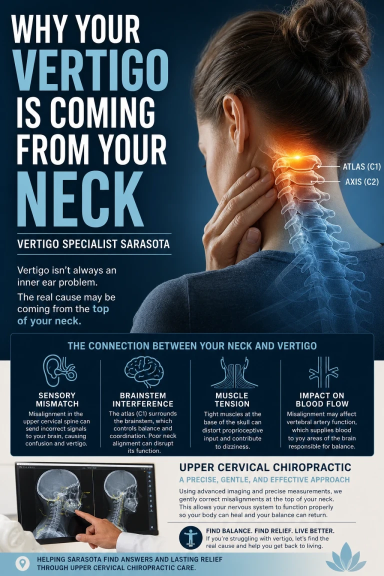
Ho and colleagues, in Head & Neck in 2015, characterised styloid and C1 transverse process juxtaposition as a cause of Eagle syndrome, reporting a series of patients with radiographic jugular compression who underwent styloid excision. In their series, styloid process lengths ranged widely, and the distance between the styloid and the C1 transverse process measured between roughly half a millimetre and under five millimetres. That is the whole corridor.
Scerrati and colleagues, reviewing this literature in Annals of Translational Medicine in 2021, described extrinsic compression of the jugular vein at the passage between the C1 transverse process and the styloid process as an emerging entity, noting that internal jugular vein stenosis has been reported in association with conditions including Ménière disease, idiopathic intracranial hypertension, and multiple sclerosis. A 2019 series in BMC Neurology reviewing twenty-three patients with symptomatic styloid elongation found the jugular variant clinically distinct from the classic and carotid forms.
One refinement is worth knowing. A morphometric study using three-dimensional CT analysis found that patients with styloid jugular nutcracker had a more vertically directed styloid process, and that this orientation mattered more than the absolute distance between the two bones. Length alone is not the whole story. Direction and spatial relationship are.
What impaired venous outflow feels like
If blood cannot leave the head efficiently, pressure inside the skull rises. The reported symptom picture includes headache, tinnitus, dizziness, visual disturbance including double vision, a sensation of pressure behind the eyes, nausea, neck pain, and papilloedema on examination. In severe cases, cerebral venous sinus thrombosis has been reported.
This list overlaps heavily with idiopathic intracranial hypertension, and distinguishing the two matters because the treatments differ entirely. The literature offers useful discriminators: compared with idiopathic intracranial hypertension, patients with styloidogenic jugular compression showed lower rates of obesity, higher rates of positional headache, longer styloid processes, and a shorter distance between the styloid and the C1 lateral tubercle.
Positional headache is the detail worth holding onto. A headache that changes clearly with head position or rotation is a mechanical signal, and it is the kind of history that gets dismissed as unremarkable when nobody is listening for it.
Related article

Sub-occipital neuralgia Treatment Sarasota, Venice, Bradenton and Tampa Bay
Nov 29, 2021Where upper cervical care fits, and where it does not
I want to be exact about this, because it would be easy to overstate and the consequences of overstating it here are more serious than usual.
An elongated styloid process is a bone. No adjustment shortens it. Symptomatic vascular Eagle syndrome is managed surgically — styloidectomy by transoral or transcervical approach, sometimes with resection of the C1 transverse process tubercle, and in some cases venous stenting. Upper cervical chiropractic is not a treatment for Eagle syndrome, and there is no trial evidence that it relieves styloidogenic compression.
What can be said is narrower and still useful. The C1 transverse process is one of the two structures forming the compression, and the position of the atlas determines where that transverse process sits. Rotation and lateral displacement of the atlas alter the geometry of a corridor already measured in fractions of a millimetre. That is a mechanically coherent proposition, and I am labelling it as a reasoned hypothesis rather than a demonstrated mechanism, because no study has shown that changing atlas position changes jugular flow in these patients.
The genuine contribution an upper cervical practice makes to this condition is identification. Patients with Eagle syndrome are frequently misdiagnosed for years — carrying labels of atypical facial pain, various neuralgias, vestibular migraine, or idiopathic intracranial hypertension — because nobody imaged the styloid process or measured its relationship to the atlas. Anyone whose headache, tinnitus, dizziness, or visual symptoms change with head rotation deserves to have that corridor looked at.
A safety point I want to state plainly
If an elongated styloid process is impinging on the carotid artery, rotational manipulation of the neck is a bad idea. Case reports describe arterial injury and pseudoaneurysm from styloid contact with the internal carotid artery, and forceful rotation is precisely the movement that changes the relationship between the styloid and the vessel.
Upper cervical correction as practised here involves no twisting, no popping, no cracking, and no pulling of the neck, which removes the specific mechanism of concern. But the more important point is the one before that: suspected carotid involvement is a reason for vascular imaging and specialist referral, not for a course of manual care of any kind. Anyone offering to adjust their way through it has not understood the risk.
Related article
Lingering Concussion Symptoms? Sarasota Upper Cervical Chiropractor Explains a Different Approach
Apr 07, 2026Imaging and objective testing
Three-dimensional imaging is what makes this diagnosis possible, and it is why the styloid process is so often missed on plain films and standard views. Cone beam computed tomography images the styloid process, the stylohyoid ligament, and the transverse process of the atlas in three dimensions, allowing the length, orientation, and spatial relationship to be measured against your own anatomy rather than a population average. Where the images raise concern about the carotid artery or the jugular vein, CT angiography, CT venography, or duplex ultrasonography with a vascular specialist is the appropriate next step. Cone beam imaging measures bone geometry. It does not image flow.
Alongside imaging, objective measures are taken at every visit: leg length assessment, cervical range of motion, postural and balance measurement, and paraspinal thermography to track whether an individual's pattern has stabilised. Those measures determine whether a correction is indicated at all. When they show the correction is holding, no adjustment is given. Repeatedly adjusting a spine that is already holding adds mechanical input without benefit, and in a region where a millimetre of geometry matters, restraint is part of the technique.
Red flags
Sudden severe headache, sudden visual loss, weakness or numbness on one side of the body, facial droop, or slurred speech requires emergency evaluation. Progressive visual change, swelling of the optic disc found on eye examination, persistent vomiting with headache, or headache that is clearly worse when lying flat all warrant urgent medical assessment for raised intracranial pressure. Neck pain with fainting or near-fainting on head turning needs vascular evaluation before anything else. Difficulty swallowing that is worsening, unexplained weight loss, or a mass in the neck requires medical work-up.
Getting evaluated in Sarasota
If you have head pressure, tinnitus, dizziness, throat or ear pain, or visual symptoms that change with head position, and no one has ever imaged your styloid process or measured its relationship to your atlas, that is a reasonable gap to close. An upper cervical evaluation with three-dimensional imaging can identify the anatomy and, where the findings warrant it, refer you to the specialist who can actually treat it. This works alongside your physician, and for vascular findings it defers to them entirely. To arrange a consultation, visit NeckWise.
Dr. Drew Hall, Upper Cervical Chiropractor
Serving Sarasota, Bradenton, and Lakewood Ranch, Florida
This article is for general educational purposes only and is not medical advice, a diagnosis, or a treatment recommendation for any individual. Eagle syndrome should be diagnosed and managed by qualified medical providers, and the vascular variants involving the carotid artery or internal jugular vein require specialist evaluation and are treated surgically or endovascularly. Upper cervical chiropractic is an area of focus within chiropractic; it is not a board-recognized specialty, and no claim of specialization or superiority is made or implied. Upper cervical chiropractic care is not a treatment for Eagle syndrome and cannot alter an elongated styloid process; no such claim is made here, and no trial evidence exists for chiropractic care in this condition. The relationship described between atlas position and the styloid-to-C1 corridor is presented as a reasoned anatomical hypothesis, not a demonstrated mechanism. Suspected carotid artery involvement is a contraindication to rotational cervical manipulation and requires vascular imaging and specialist referral. Cone beam computed tomography is used as a precision measurement and analysis tool, not as a stand-alone diagnostic device, and does not assess blood flow. Individual results vary and no specific outcome is guaranteed. Seek emergency medical attention for sudden severe headache, sudden visual loss, weakness or numbness on one side of the body, facial droop, or slurred speech. Always consult a qualified healthcare provider regarding diagnosis and treatment.


Leave a comment