

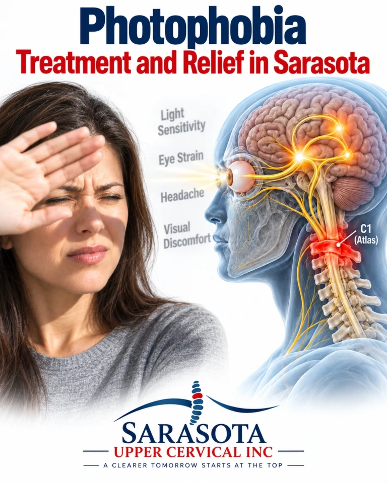
Light sensitivity, or photophobia, is treated as a symptom rather than a problem in Sarasota, Bradenton, and Lakewood Ranch — something mentioned in passing while discussing migraine or concussion and rarely investigated on its own. It deserves better, because the pathway that produces it is well mapped, and it runs directly through the same brainstem and upper cervical structures that process head and neck pain.
Photophobia is not sensitive eyes. It is a pain pathway being triggered by light.
Schedule Your Appointment
Schedule appointmentWhat photophobia actually is
The word implies fear of light, which is misleading. Clinically it describes light of ordinary intensity producing discomfort or frank pain, along with squinting, aversion, watering, and often worsening of an underlying headache.
The critical observation, and the one that reframes the whole condition, is that photophobia can occur in people who are blind. Patients without functional image-forming vision — who cannot perceive light consciously — can still experience light-induced discomfort. That rules out the visual pathway as the mechanism and points to something else.
The mechanism A separate class of retinal cell
The retina contains rods and cones, which serve image formation, and a third and much smaller population: intrinsically photosensitive retinal ganglion cells containing the pigment melanopsin. These cells respond to light directly, independent of rods and cones, and they do not contribute to seeing. They project instead to non-image-forming targets, including the circadian clock in the hypothalamus, the pupillary reflex pathway, and — critically here — the thalamus.
This is why photophobia persists in some blind patients whose retinal ganglion cells remain intact even when the image-forming pathway does not.

Convergence with dural pain
The pivotal finding, established through work by Rami Burstein and colleagues, is that these light-responsive signals converge in the posterior thalamus onto the same neurons that receive nociceptive input from the dura via the trigeminal system.
That convergence is the mechanism. A thalamic neuron receiving both light-driven and pain-driven input cannot separate them. Light arriving at a neuron already carrying dural nociceptive traffic increases its firing. The result is that light is experienced as pain, and that light makes an existing headache worse.
There is also a trigeminal-autonomic component. Trigeminal fibres innervating the eye and its blood vessels, and reflex connections through the superior salivatory nucleus, account for the tearing, redness, and ocular ache that accompany severe photophobia.
Where the upper cervical spine enters
Trigeminal nociceptive fibres from the dura and cranial vessels do not synapse at the pons. They descend as the spinal trigeminal tract through the medulla and terminate in the trigeminal nucleus caudalis, which extends into the upper cervical spinal cord to approximately C2 and often C3.
Afferents from the C1, C2, and C3 nerve roots enter at those same levels and synapse on the same second-order neurons — the trigeminocervical complex. Many of these are wide dynamic range neurons integrating input across a broad receptive field, and they cannot distinguish dural input from suboccipital input.
Follow that forward. Second-order neurons in the trigeminocervical complex project to the thalamus, where the convergence with light-driven signals occurs. Upper cervical afferent traffic therefore feeds into the same ascending pathway that photophobia depends on. Raising the background activity of the trigeminocervical complex plausibly lowers the threshold at which light becomes painful.
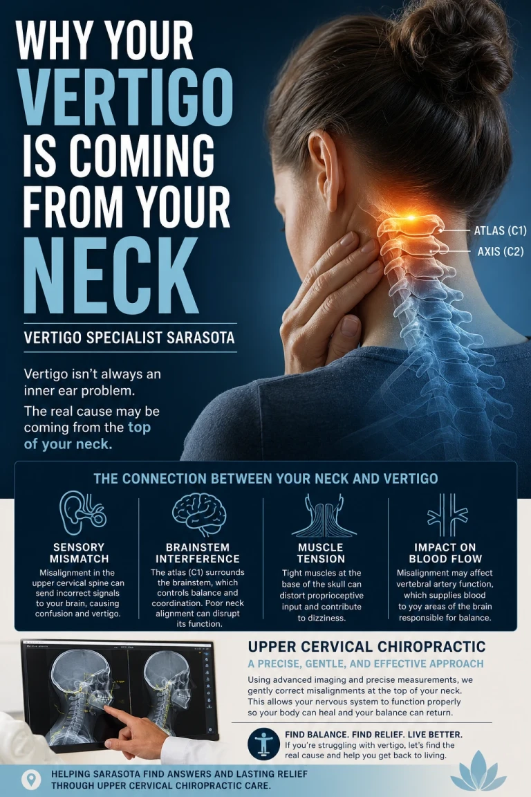
I want to mark the boundary clearly. Every link in that anatomical chain is established: the melanopsin cells, the thalamic convergence, the descent of the trigeminal nucleus into the upper cervical cord, the convergence of cervical afferents there. What has not been demonstrated is that reducing upper cervical input measurably raises photophobia thresholds in humans. That step is a reasoned inference, and there is no clinical trial of upper cervical chiropractic for photophobia.
The conditions photophobia accompanies
Migraine is the most common association, and photophobia is part of the diagnostic criteria. Many migraine patients report a persistent lower light tolerance even between attacks, consistent with a chronically sensitised pathway rather than an attack-only phenomenon.
Post-concussion syndrome frequently includes photophobia, often alongside convergence problems and difficulty with visually busy environments. Since concussion mechanisms almost invariably load the cervical spine as well as the brain, disentangling the cervical contribution matters.
Blepharospasm, dry eye, uveitis, corneal disease, and acute glaucoma all produce light sensitivity from the eye itself and require ophthalmological assessment. Meningitis produces photophobia with fever and neck stiffness and is a medical emergency. Trigeminal neuralgia, thalamic lesions, and some medications also appear on the list.
What to do about the light itself
One practical point deserves emphasis because it is counterintuitive and frequently gotten wrong.
Sustained dark adaptation makes photophobia worse. Wearing dark glasses indoors or living in a dimmed room lowers the threshold further over time, and the condition becomes self-reinforcing. Guidance in the headache literature discourages habitual indoor dark lens use for this reason.
What appears more useful is addressing the specific wavelengths and flicker characteristics that drive the response — reducing exposure to fluorescent flicker, using precision tinted lenses where indicated by an appropriate assessment rather than generic dark lenses, and adjusting screen brightness to match ambient light rather than exceeding it. Gradual, graded reintroduction of normal lighting, rather than avoidance, follows the same logic as graded exposure in any sensitised system.
Precision and knowing when not to adjust
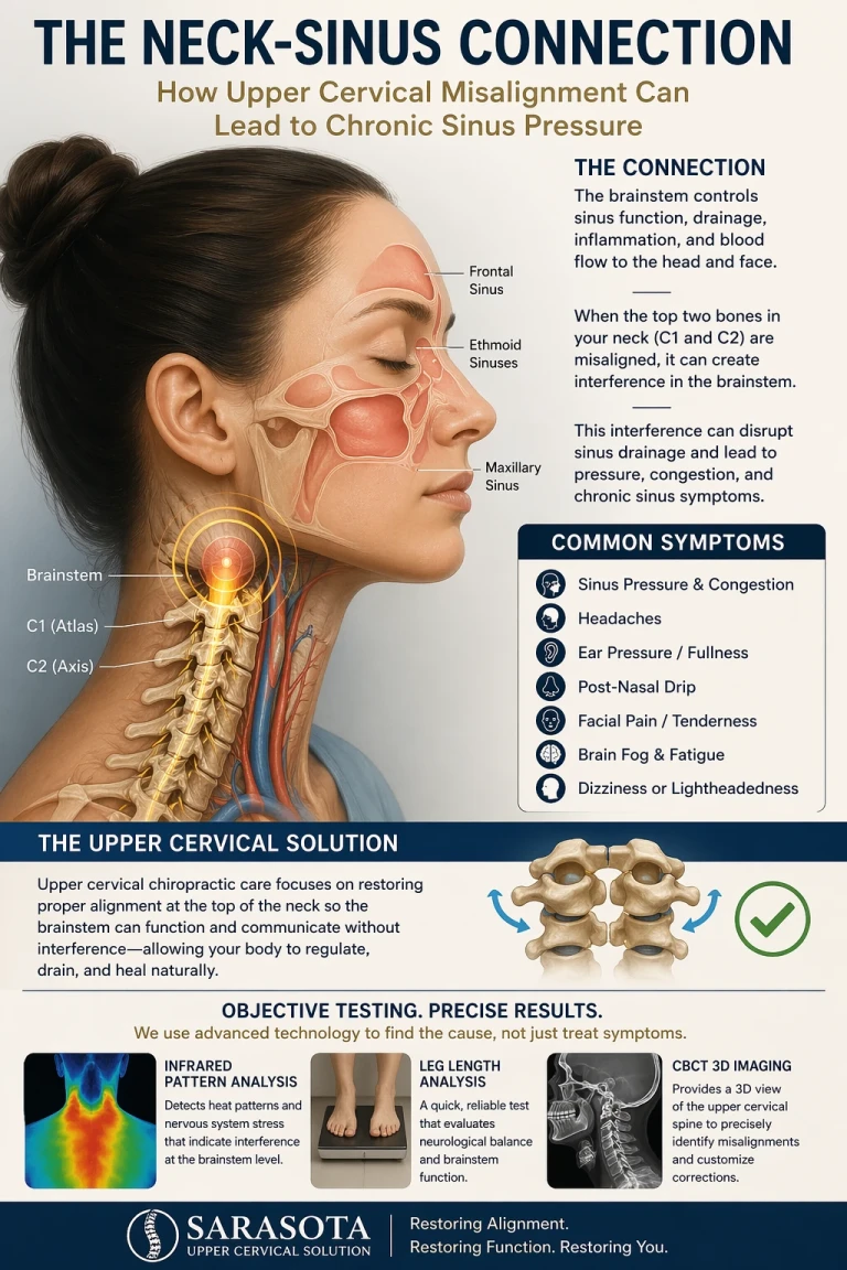
Upper cervical correction involves no twisting, no popping, no cracking, and no pulling of the neck. Cone beam computed tomography measures the three-dimensional position of the atlas and axis against your own anatomy so a correction is calculated rather than estimated. Objective measures — leg length assessment, cervical range of motion, postural and balance measurement, and thermographic pattern tracking — determine whether a correction is indicated and whether it is holding. When it is holding, no adjustment is given.
In a sensitised trigeminal system that restraint is the point rather than a formality. Additional input into an already overloaded pathway is not neutral, and the clinic environment itself should account for the condition — a patient with severe photophobia should not be assessed under bright fluorescent lighting.
Red flags requiring urgent evaluation
Photophobia with fever and neck stiffness requires emergency evaluation for meningitis. A red painful eye with light sensitivity, blurred vision, and haloes around lights may indicate acute angle-closure glaucoma or uveitis and requires same-day ophthalmological assessment. Sudden severe headache with photophobia reaching maximum intensity within seconds requires emergency assessment. New photophobia with visual loss, double vision, weakness, numbness, or confusion requires urgent neurological evaluation. Photophobia following head trauma warrants medical assessment. Unilateral photophobia with eye pain is more likely ocular than neurological and needs examination.
An evaluation in Sarasota
If your eyes have been examined and are healthy, and light still hurts — particularly alongside headache, neck pain, or symptoms that began after a concussion or whiplash injury — an upper cervical evaluation can assess whether cervical input is contributing to a sensitised trigeminal pathway. This works alongside your eye doctor, neurologist, and headache care rather than in place of it, and an eye examination should come first. To arrange a consultation by calling 941 259-1891.
Dr. Drew Hall, Upper Cervical Chiropractor
Serving Sarasota, Bradenton, and Lakewood Ranch, Florida
This article is for general educational purposes only and is not medical advice, a diagnosis, or a treatment recommendation for any individual. Light sensitivity requires examination by a qualified optometrist or ophthalmologist to exclude ocular causes, and medical evaluation where a neurological cause is suspected. Upper cervical chiropractic is an area of focus within chiropractic; it is not a board-recognized specialty, and no claim of specialization or superiority is made or implied. No clinical trial evidence exists for upper cervical chiropractic care in photophobia, and none is claimed here; the melanopsin and thalamic convergence pathway described is established physiology, and the proposed contribution of upper cervical afferent input to photophobia thresholds is a reasoned inference rather than a demonstrated mechanism. Cone beam computed tomography is used as a precision measurement and analysis tool, not as a stand-alone diagnostic device. Individual results vary and no specific outcome is guaranteed. Seek emergency care for photophobia with fever and neck stiffness, sudden severe headache, a red painful eye with haloes or visual loss, or photophobia with weakness, numbness, double vision, or confusion. Always consult a qualified healthcare provider regarding diagnosis and treatment.



Leave a comment