

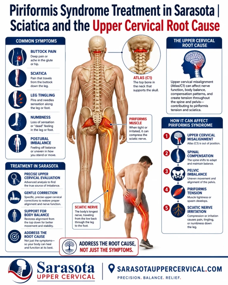
Piriformis syndrome treatment usually begins and ends at the piriformis muscle itself, which is a reasonable place to start and an incomplete place to stop. Buttock pain that radiates down the back of the leg, worsens with sitting, and fails to resolve with stretching and soft tissue work is common enough in Sarasota that most people who have it have already tried the standard approach. This article makes a specific argument about why that approach sometimes stalls, what the upper cervical spine has to do with resting muscle tone in the pelvis, and where the evidence for that argument is strong, where it is suggestive, and where it is honestly still a hypothesis.
What Piriformis Syndrome Is, and Why the Diagnosis Is Contested
Schedule Your Appointment
Schedule appointmentThe piriformis originates on the anterior surface of the sacrum around the S2 to S4 levels, passes out through the greater sciatic notch, and attaches to the greater trochanter of the femur. In the majority of people the sciatic nerve exits the pelvis directly beneath it. The two structures share a tight corridor, and when the muscle is held short and firm, that corridor narrows.
The condition is difficult to pin down. Published estimates of how much sciatica piriformis syndrome accounts for run from well under one percent to more than a third of cases depending on the source, which tells you the diagnostic criteria are not settled. A 2025 systematic review of 212 published cases found that 38.2 percent of patients had a history of blunt or indirect pelvic trauma or vigorous physical stress on the muscle, and that in 47.6 percent of cases no instrumental or intraoperative finding was ever identified to explain the presentation. That last number matters. In nearly half of documented cases, nothing structural was found. Something functional was driving it.
It is also worth noting that the case report literature skews heavily toward severe and refractory presentations, since routine cases that resolve do not get written up. Conclusions drawn from it should be read with that bias in mind.
Why Stretching Helps, and Why It Often Stops Helping
Piriformis stretches are worth doing, and this article is not an argument against them. Supine figure-four positions, seated piriformis stretches, hip external rotator work, glute medius strengthening, and postural correction for prolonged sitting all have a legitimate role. Some people recover completely with nothing more than that. Manual therapy, post-isometric relaxation, myofascial release, and targeted strengthening of the hip abductors and external rotators have small trials supporting them, though the trial base is heterogeneous and generally low in methodological quality.
The pattern worth paying attention to is the one where stretching produces real but temporary relief. The muscle lengthens, the symptoms ease for a day or two, and then the tightness returns without any obvious provocation. That pattern has a straightforward interpretation. Stretching changes muscle length. It does not change the neural drive setting resting tone in that muscle. If something upstream is continuously instructing the piriformis to hold at a shortened resting length, then stretching is treating an output while the input keeps running.
Which raises the question that most discussions of this condition skip entirely. Muscles do not set their own resting tone. So where is the tone of the piriformis actually coming from?
Postural Muscle Tone Is Regulated From the Top of the Spine
Resting tone in postural muscles is an output of brainstem circuitry, delivered through the vestibulospinal and reticulospinal tracts, and continuously updated by sensory information about where the head sits relative to the body. The densest single source of that information is the upper cervical spine.
The suboccipital muscles carry an extraordinary concentration of muscle spindles, with reported densities as high as roughly 200 per gram of tissue compared with about 16 per gram in the first lumbrical of the hand. These are not muscles built for strength. They are built to report head position at very high resolution. That signal feeds the cervicocollic reflex, the cervico-ocular reflex, and the tonic neck reflex, and the tonic neck reflex integrates with vestibulospinal output directed at trunk and limb muscles.
The experimental demonstration is direct. Vibrating the posterior neck muscles in healthy adults produces short-latency EMG activation in lower leg muscles and measurable postural reactions at the feet. Investigators controlled for the possibility that vibration was simply exciting the nearby vestibular organs by applying the same stimulus over the mastoid, and cervical afferents still dominated the response. Earlier work showed neck input modulating the soleus H-reflex, meaning cervical signals reach into the leg and alter spinal reflex excitability there.
A pathway from cervical proprioceptive input to lower limb motor output is not speculative. It is measurable in a laboratory. The clinical question is what happens to that pathway when the upper neck has been injured.
Mechanism One: Aberrant Proprioceptive Input After Neck Injury
Work from Winkelstein's group at the University of Pennsylvania and Panjabi's group at Yale established that the cervical facet capsular ligament is injured during whiplash-type loading well below the threshold at which it visibly fails. Panjabi and colleagues estimated capsular strains in the range of thirty to forty percent during simulated whiplash, and minor ruptures have been documented at strains between roughly thirty-five and sixty-five percent. Subfailure loading of this kind increases joint laxity and alters ligament stiffness without producing anything a standard radiograph would call abnormal.
What lives in that capsule is the important part. It is densely innervated with both proprioceptive and nociceptive mechanoreceptors, whose peripheral terminals sit in the capsule itself. Injuring it does not simply create a painful joint. It damages a sensor. And when a segment subsequently loses normal motion, whether from guarding, adhesion, or altered mechanics, the mechanoreceptors in that segment stop receiving the movement they are built to report on. The signal reaching the brainstem is not merely reduced. It is wrong.
This is measurable in patients. Cervical joint position error testing asks a person to return their head to a neutral starting position with eyes closed after rotating away from it. Revel and colleagues established in 1991 that healthy controls typically return within about four and a half degrees. Sterling and colleagues reported in 2003 that a whiplash group averaged just over five degrees of error against 1.75 degrees in healthy controls. Elevated joint position error is a documented finding in whiplash-associated disorder and chronic neck pain, and it is generally interpreted as evidence of mismatched cervical afferent input rather than as a problem with the neck muscles themselves.
The brainstem has no independent way of verifying that the position signal it is receiving is inaccurate. It builds a postural strategy on the information available, and it commits the rest of the frame to that strategy.
Mechanism Two: The Myodural Bridge and Dural Tension
There is a second anatomical feature of the suboccipital region that deserves attention. In 1995, Hack and colleagues described a dense fibrous connection running from the rectus capitis posterior minor muscle through the posterior atlanto-occipital membrane to the spinal dura mater, and named it the myodural bridge. Subsequent work has confirmed similar connections involving the rectus capitis posterior major, the obliquus capitis inferior, and the ligamentum nuchae. In one histological study, all twelve obliquus capitis inferior specimens examined emitted grossly visible soft tissue tracts inserting into the posterolateral cervical dura, and staining identified neuronal fascicles traveling within the bridge itself.
The anatomy is well established at this point, confirmed through gross dissection, histology, plastinated cross-section, and MRI across multiple independent groups. The proposed functions are less settled. Researchers have suggested the bridges prevent the dura from infolding, help maintain patency of the cerebellomedullary cistern, contribute to cerebrospinal fluid circulation, and transmit dural tension. These remain hypotheses supported by anatomy rather than conclusions established by experiment.
The clinically relevant idea is this. If suboccipital muscles enter sustained protective tone after a neck injury, that tone has a mechanical destination beyond the muscle. It reaches the dura. And the dura is a continuous sleeve running the entire length of the spinal canal to its anchoring points at the sacrum and coccyx. A tension change introduced at the craniocervical junction is not confined there. Whether that tension is sufficient to influence lumbosacral neural mechanics in a way that matters clinically has not been demonstrated, and anyone presenting it as settled is going beyond what the literature supports. It is a plausible additional pathway, worth stating and worth labeling honestly.
The Chain Reaction Down the Spine
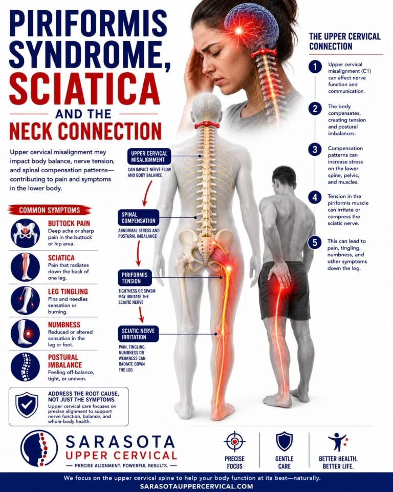
The nervous system holds the eyes level and the head upright at almost any cost to the rest of the frame. If the top of the spine reports a head tilt that is not present, or fails to report one that is, the compensation does not stay local. Postural tone gets redistributed asymmetrically downward through the thoracic spine, the lumbar spine, and the pelvis to keep the head where the brainstem believes it should be.
Pelvic torsion produced this way is a tone phenomenon rather than a bone phenomenon. It shows up on examination as an apparent leg length asymmetry with the patient lying down, which reviews of the topic attribute to hypertonicity in muscles above the pelvis rather than to any actual difference in bone length. The piriformis sits directly in the middle of that compensation. It is small, deep, postural, attached to the sacrum on one end and the femur on the other, and it shares a narrow corridor with the largest nerve in the body. It does not take much sustained shortening for that corridor to become symptomatic.

What the Research Actually Shows, and What It Does Not
The most directly relevant study is Nansel and colleagues, published in the Journal of Manipulative and Physiological Therapeutics in 1993. In a randomized, double-blind before-and-after design, adjustments were delivered to either the upper cervical spine at C2 or the lower cervical spine at C7, and lumbar paraspinal tissue compliance was measured at the L1 through L5 levels before and within fifteen minutes afterward. Cervical adjustments produced significant changes in lumbopelvic muscle tone, which the authors attributed to facilitation of tonic neck reflexes through intersegmental spinal pathways.
That is a real finding and it is directly on point. It is also a small study in healthy asymptomatic chiropractic students, measuring an immediate effect with a tissue compliance meter, with no symptomatic population and no follow-up. It demonstrates that the pathway is active. It does not demonstrate a clinical outcome.
Honesty requires the counterweight as well. A 2012 systematic review of spinal manipulative therapy and range of motion found no effect of manipulation on lumbar range of motion across three studies, and a study of sacroiliac manipulation reported no effect on hip range of motion. The literature on manipulation and remote biomechanical change is genuinely mixed, and a reader deciding what to do should know that.
The clearest statement of the current position is this. No study has demonstrated that an upper cervical misalignment causes piriformis hypertonicity, and no clinical trial has tested upper cervical chiropractic care as a treatment for piriformis syndrome. The physiology connecting cervical afferent input to lower limb postural tone is established. The extension of that physiology to this specific muscle and this specific pain pattern is a reasoned clinical hypothesis under test, not a proven mechanism.
Cone Beam CT and the Case for Precision
Upper cervical anatomy varies substantially between individuals. Occipital condyle shape, atlas symmetry, the odontoid relationship, and developmental variants such as ponticulus posticus differ enough from person to person that a correction vector calculated from assumed anatomy is a correction vector calculated from a guess. Cone beam CT resolves the actual anatomy in three dimensions rather than requiring it to be inferred from a two-dimensional projection.
Its role is narrower than it is sometimes made out to be. Cone beam imaging does not diagnose piriformis syndrome. It does not image muscle tone, it does not image the sciatic nerve, and it says nothing about the pelvis. What it does is establish what the individual anatomy is, identify findings that would make adjusting inappropriate or require modification, and allow a correction to be planned specifically rather than generically. It is also ionizing radiation, at a dose lower than conventional medical CT but higher than plain film, which means it should be ordered because a clinical question requires it and not as routine.
Objective Testing, and Knowing When Not to Adjust
Objective pre-checks are performed at every visit before any decision to adjust is made. If the indicators say the previous correction is holding, no adjustment is delivered that day. Post-checks are performed after any correction to verify that something measurably changed rather than assuming it did.
This matters more than it initially sounds. A practice that adjusts on a schedule rather than on findings has stopped testing and started performing. Repeatedly applying force to a joint complex that is already stable does not improve on stable, and in a patient whose capsular tissue has already been injured, it delivers mechanical insult to tissue that is trying to repair.
Why Holding the Correction Is the Point
Ligament healing follows a well-characterized sequence. An inflammatory phase occupies roughly the first week. A proliferative phase spans the following weeks, during which fibroblasts lay down disorganized type III collagen that is mechanically weaker than the original tissue. A remodeling phase then runs from around six weeks out to twelve months or considerably longer, during which type III collagen is progressively replaced by stronger, better-aligned type I collagen and the fibers reorganize along lines of mechanical stress.
Two features of that process are important here. The first is that remodeled ligament is not restored ligament. The literature is consistent that repaired ligament tissue is morphologically and biomechanically inferior to native tissue, with reduced strength and durability, which is why residual laxity after ligamentous injury is common. The second is that the remodeling phase is genuinely vulnerable. Controlled mechanical loading during remodeling helps collagen align correctly. Excessive or repeated disruption during the same window degrades the result.
The rationale for the upper cervical approach follows from those two facts rather than from anything unique to chiropractic. If the capsular tissue at the craniocervical junction was injured, and if the segment is corrected and then left alone while it remodels, the tissue has an uninterrupted window in which to consolidate. As stability improves, the mechanoreceptors in that capsule return to reporting on more normal motion, the afferent picture reaching the brainstem improves, and the postural tone strategy built on the old, corrupted picture has an opportunity to settle. A more balanced distribution of postural tone is what would eventually reach the piriformis.
That chain is coherent and it is grounded in documented tissue physiology at every link. It has not been demonstrated end to end by a clinical trial. No study has tested whether adjusting frequency affects cervical capsular ligament healing, and no study has tracked piriformis tone as an outcome of upper cervical care. Presented as an established mechanism it would be overselling. Presented as the reasoning behind a conservative, test-driven approach, it is defensible.
What Recovery Realistically Looks Like
If the tissue timeline above is the operative one, then the honest expectation is measured in months rather than visits, and progress is not linear. Some people notice change in postural signs before they notice change in symptoms. Some notice symptom change first. Some do not respond at all, which is information rather than failure, and is a reason to reconsider the working hypothesis rather than to continue indefinitely.
Local work on the piriformis is not abandoned during this. Stretching, hip external rotator and abductor strengthening, and sitting modification remain useful, and in many cases they become more durable once the drive maintaining the tone has been addressed. The argument here is about sequence and emphasis, not about replacing one thing with another.
When Upper Cervical Care Is Not the Right Answer
Certain findings require emergency medical evaluation rather than conservative care of any kind. New bowel or bladder dysfunction, numbness in the saddle region, and progressive or bilateral leg weakness may indicate cauda equina syndrome and warrant immediate hospital assessment. Progressive foot drop, unexplained weight loss, a history of cancer, fever, or significant recent trauma all require medical workup first.
Upper cervical adjusting is itself contraindicated or requires substantial modification in the presence of rheumatoid arthritis, Down syndrome, connective tissue disorders such as Ehlers-Danlos, os odontoideum, known craniocervical instability, active anticoagulation, and suspected vertebrobasilar insufficiency. High-velocity manipulation is specifically inappropriate in the presence of frank instability. Imaging exists in part to identify these before anything is done.
Piriformis syndrome also coexists with other conditions more often than it excludes them. Lumbar disc herniation, foraminal stenosis, sacroiliac dysfunction, hip pathology, and deep gluteal syndrome arising from structures other than the piriformis can all produce overlapping presentations, and more than one can be present at once. Any evaluation that arrives at a single answer quickly should be viewed with suspicion, and a patient who is not responding deserves reassessment rather than more of the same.
Piriformis Syndrome Care in Sarasota, Bradenton, and Lakewood Ranch
Buttock and leg pain that has not responded to treatment aimed locally at the piriformis is worth examining from further up the chain, particularly when there is a history of head or neck trauma, whiplash, concussion, or a significant fall, and particularly when stretching produces relief that reliably fades. The examination process, the imaging, the objective indicators used to decide whether an adjustment is appropriate on a given day, and what a realistic course of care involves.
Written by Dr. Drew hall, Upper Cervical Chiropractor.
This article is provided for general educational purposes only and does not constitute medical advice, diagnosis, or treatment, and it does not establish a doctor-patient relationship. Individual results vary and no specific outcome is guaranteed. Do not use this information to diagnose or treat a health problem or to delay seeking care from a qualified healthcare provider. If you are experiencing new bowel or bladder changes, saddle numbness, progressive leg weakness, or any other symptom you believe may be an emergency, seek immediate medical attention.


Leave a comment