

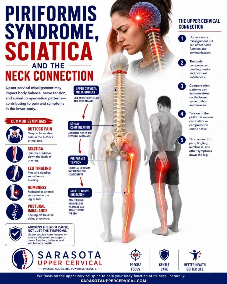
Piriformis syndrome describes buttock and posterior leg pain believed to arise when the piriformis muscle compresses or irritates the sciatic nerve, and it is one of the most contested labels in musculoskeletal medicine. Published estimates of how much sciatica it accounts for range from under one percent to more than a third of cases depending on which paper you read. That spread says less about the condition than about the absence of agreed diagnostic criteria, and it is worth stating before proposing any explanation for it, including the one this article makes.
The Muscle, the Nerve, and the Space They Share
Schedule Your Appointment
Schedule appointment
The piriformis originates on the anterior sacrum around the S2 to S4 levels, passes through the greater sciatic notch, and attaches to the greater trochanter. In most people the sciatic nerve exits the pelvis immediately beneath it, and the two structures share a small, crowded corridor.
The conventional explanation leans heavily on anatomical variation. Beaton and Anson described six possible relationships between the sciatic nerve and the piriformis in 1938, and their classification is still standard. Roughly eighty-seven percent of people have the conventional arrangement, an undivided nerve running beneath an intact muscle. The remaining thirteen percent have some variant, most often the common peroneal division passing through a split muscle. The intuitive conclusion is that the variant group is the one that develops entrapment.
The Anatomy Argument Has a Problem
That conclusion does not survive contact with the imaging data. A 2018 study in European Radiology reviewed 783 consecutive adult hip MRIs, classified each by Beaton and Anson type, and cross-referenced charts for the clinical diagnosis of piriformis syndrome. Variants were present in 19.2 percent of hips. There was no statistically significant difference in the prevalence of piriformis syndrome, sciatica, or buttock pain between the variant and conventional groups.
This is one retrospective study and not the final word. But it points somewhere important. If the structural arrangement of nerve and muscle is not what separates the people who hurt from the people who do not, then something about the behavior of the muscle has to be doing the separating. The most obvious candidate is resting tone. A piriformis at normal length does not compress much of anything, variant anatomy or not. One held chronically short narrows an already narrow space.
Which raises the question most discussions of this condition skip. Nothing sets its own tone. So where is the tone of the piriformis actually being set?
Where Postural Muscle Tone Is Regulated
Related article
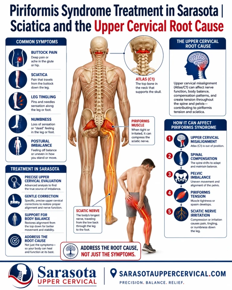
Piriformis Syndrome Treatment in Sarasota | Sciatica and the Upper Cervical Root Cause
Aug 13, 2026
Resting tone in postural muscles is not a local property. It is an output of brainstem circuitry delivered through the vestibulospinal and reticulospinal tracts, continuously updated by sensory information about where the head sits relative to the body. The densest single source of that information is the upper cervical spine.
The suboccipital muscles carry an extraordinary concentration of muscle spindles, with reported densities as high as roughly 200 per gram of tissue compared with about 16 per gram in the first lumbrical of the hand. These are not muscles built for strength. They are built to report head position at high resolution. That signal feeds the cervicocollic reflex, the cervico-ocular reflex, and the tonic neck reflex, and the tonic neck reflex integrates with vestibulospinal output directed to trunk and limb muscles.
The experimental demonstration is direct. Vibrating the posterior neck muscles in healthy adults produces short-latency EMG activation in lower leg muscles and measurable postural reactions at the feet. Investigators controlled for the possibility that vibration was merely exciting the nearby vestibular organs by applying the same stimulus over the mastoid, and cervical afferents still dominated. Earlier work showed neck input modulating the soleus H-reflex, meaning cervical signals alter spinal reflex excitability in the leg. A real, measurable pathway runs from cervical proprioceptive input to lower limb motor output. What follows is where honesty matters most.
What Has Not Been Shown
No study has demonstrated that an upper cervical misalignment causes piriformis hypertonicity, and no clinical trial has tested upper cervical chiropractic care as a treatment for piriformis syndrome. The link between cervical afferent input and lower limb tone is well established under laboratory conditions. Extending that finding to this specific muscle and this specific pain pattern is a reasoned hypothesis, not a proven mechanism, and anyone presenting it otherwise is overselling.
How a Neck Injury Becomes a Whole-Spine Problem
The trauma piece is better supported. Work from Winkelstein's group at Penn and Panjabi's group at Yale established that the cervical facet capsular ligament is injured during whiplash-type loading well below the threshold at which it visibly fails. Panjabi and colleagues estimated capsular strains around thirty to forty percent in simulated whiplash, and minor ruptures have been documented between roughly thirty-five and sixty-five percent strain. Subfailure loading of this kind increases joint laxity and alters stiffness without producing anything a standard radiograph would call abnormal.
The important detail is what lives in that capsule. It is densely innervated with both proprioceptive and nociceptive mechanoreceptors. Injuring it does not simply create a painful joint. It corrupts a sensor, and the brainstem has no way of knowing it is now working from bad data.
Related article
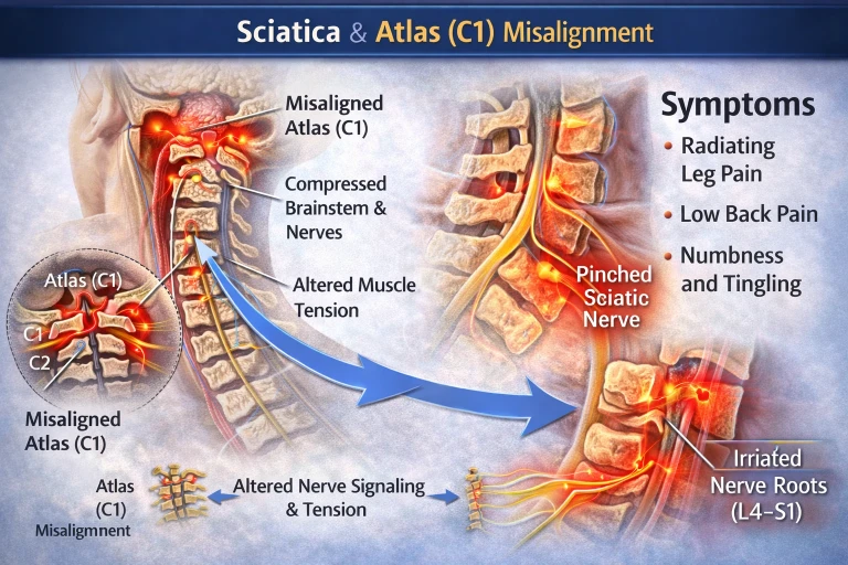
Sciatica Treatment in Sarasota, Bradenton & Lakewood Ranch | Chiropractor Near Me
Mar 09, 2026From there the cascade follows from priority. The nervous system will keep the eyes level and the head upright almost regardless of what it costs the rest of the frame. If the top of the spine reports a tilt that is not there, compensations get distributed downward through the thoracic spine, lumbar spine, and pelvis as sustained asymmetric tone. The piriformis is a plausible casualty. It is small, deep, postural, and it shares a tight corridor with the largest nerve in the body. It does not take much sustained shortening to matter.
What Cone Beam CT Actually Contributes
Cone beam CT of the upper cervical spine does one thing well. It shows individual anatomy in three dimensions rather than inferring it from a flat projection, including condyle shape, atlas symmetry, the odontoid relationship, and developmental variants such as ponticulus posticus. Upper cervical anatomy varies substantially between people, and a correction vector calculated from assumed anatomy is calculated from a guess.
What it does not do is diagnose piriformis syndrome. It does not image muscle tone or the sciatic nerve and says nothing about the pelvis. Its role is narrower than it is often made out to be: establish the anatomy, rule out findings that would make adjusting inappropriate, and make any correction specific rather than generic.
Leg Checks, Described Honestly
Functional leg length asymmetry observed with the patient lying down is not a measurement of anatomic leg length inequality and should never be presented as one. Rhodes and colleagues compared prone leg checks against standing radiographs in fifty patients and found a correlation of 0.71, but in twelve percent of cases the two methods identified opposite legs as the shorter one. Cooperstein and Lucente later compared supine and prone examiners on the same subjects and found the two disagreed about which leg was short in nearly forty-two percent of cases.
Those numbers are not a defense of the test as a precision instrument. They are the reason it belongs among several crude inputs rather than standing as evidence on its own. What an unloaded asymmetry probably reflects, as reviews of the topic argue, is hypertonicity in muscles above the pelvis pulling it into torsion. It is a tone sign, not a bone measurement, and that is precisely why it is relevant here. Tracked in the same position by the same examiner over time, a change in that sign carries some information. Treated as proof, it carries none.
Adjusting Only When the Testing Says To
Objective pre-checks are performed at every visit. If the indicators say the previous correction is holding, no adjustment is delivered, and post-checks follow any correction to confirm something actually changed. Repeatedly applying force to a joint complex that is already stable does not improve on stable, and it adds mechanical insult to a region that, in a patient with a history of capsular injury, has already been insulted. A practice that adjusts on schedule rather than on findings has stopped testing and started performing.
When This Is Not the Right Answer
Certain findings require emergency medical evaluation rather than conservative care of any kind. New bowel or bladder dysfunction, saddle numbness, and progressive or bilateral leg weakness may indicate cauda equina syndrome and warrant immediate hospital assessment. Progressive foot drop, unexplained weight loss, a history of cancer, fever, or significant recent trauma all require medical workup first.
Upper cervical adjusting is itself contraindicated or requires substantial modification in the presence of rheumatoid arthritis, Down syndrome, connective tissue disorders such as Ehlers-Danlos, os odontoideum, known craniocervical instability, active anticoagulation, and suspected vertebrobasilar insufficiency. Imaging exists in part to identify these before anything is done.
Piriformis syndrome also coexists with other conditions more often than it excludes them. Lumbar disc herniation, foraminal stenosis, sacroiliac dysfunction, hip pathology, and deep gluteal syndrome from other structures can all produce overlapping presentations, and more than one can be present at once. Any evaluation that arrives at a single answer quickly should be viewed with suspicion.
The Reasonable Position
Buttock and leg pain that has not responded to treatment aimed locally at the piriformis is worth examining from further up the chain, particularly when there is a history of head or neck trauma, whiplash, concussion, or a significant fall. The mechanism connecting upper cervical function to lower limb postural tone is real at the level of physiology. Its clinical relevance to this specific condition remains a hypothesis under test. That is an honest place to stand, and a better foundation for care than certainty that has not been earned.
If you are in Sarasota, Bradenton, or Lakewood Ranch and want your upper cervical spine evaluated as part of working out a persistent sciatic pattern, you can schedule a free consult by calling 941 259-1891
Written by Dr. Drew hall.
This article is provided for general educational purposes only and does not constitute medical advice, diagnosis, or treatment. It does not establish a doctor-patient relationship. Individual results vary and no specific outcome is guaranteed. Do not use this information to diagnose or treat a health problem or to delay seeking care from a qualified healthcare provider. If you are experiencing new bowel or bladder changes, saddle numbness, progressive leg weakness, or any other symptom you believe may be an emergency, seek immediate medical attention.


Leave a comment