

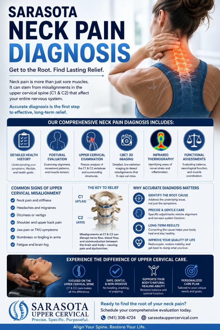
Getting neck pain properly evaluated involves more than a single X-ray glanced at in an exam room. A thorough diagnostic process moves through several layers, from history and physical exam through imaging, and the specific imaging chosen matters a great deal depending on what's actually being investigated. This guide walks through how neck pain is diagnosed step by step, including where advanced upper cervical imaging fits into a complete picture.
Step One: History and Physical Examination
The diagnostic process starts with a detailed history: how the pain started (gradually or after a specific injury), where it's located, whether it radiates into the arm, what movements worsen or ease it, and any associated symptoms such as headache, dizziness, or numbness. The physical exam then assesses range of motion, palpates for areas of tenderness or muscle guarding, and includes orthopedic and neurological tests to check for nerve involvement, reflexes, and strength.
Schedule Your Appointment
Schedule appointmentStandard X-Ray Imaging
A standard cervical spine X-ray shows the bony alignment of the vertebrae and can reveal degenerative changes such as disc space narrowing, bone spurs, and arthritic changes. Flexion-extension X-rays, taken with the neck bent forward and then backward, add an important additional layer: they can reveal abnormal movement between vertebrae, instability that a single static image would miss entirely.
MRI: Soft Tissue and Nerve Detail
When disc herniation, nerve compression, or spinal cord involvement is suspected, particularly in the presence of arm symptoms like radiating pain, numbness, or weakness, MRI provides detailed imaging of the discs, nerve roots, spinal cord, and surrounding soft tissue that X-ray cannot show.
Cone Beam CT (CBCT): Precision Imaging of the Upper Cervical Spine
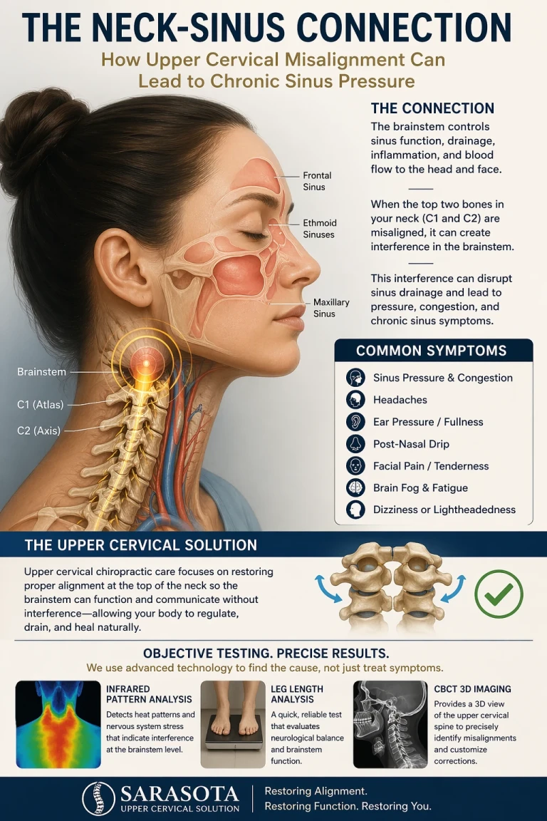
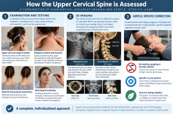
Standard X-rays are useful for general cervical alignment, but they don't provide the same level of three-dimensional precision needed to fully evaluate the atlas (C1) and axis (C2), the two vertebrae directly beneath the skull, and the segment responsible for the majority of the neck's rotational movement. Cone beam CT (CBCT) provides a detailed 3D reconstruction of this specific region, allowing precise measurement of atlas and axis position and rotation in ways a standard 2D X-ray cannot capture.
Why this level of precision matters
Because the atlas and axis sit directly beneath the brainstem, small degrees of rotational or positional change in this specific region can have a mechanical significance disproportionate to their size. The Blair upper cervical analysis method uses this kind of precise imaging specifically because generalized alignment assessment isn't sufficient at this level of the spine; the correction has to be based on each individual's actual measured anatomy, which varies from person to person.
What a Thorough Workup Looks For Beyond Bone Position
Cervical curve assessment
A lateral X-ray view allows measurement of the neck's natural forward curve. Loss of this curve, sometimes called straight neck syndrome, is a common consequence of whiplash and postural strain, and factors into the mechanical tension placed on the spinal cord and surrounding structures.
Related article

Insomnia Treatment in Sarasota | The Brainstem, Sympathetic Tone, and the Upper Cervical Connection
Aug 13, 2026Functional and neurological assessment
Beyond imaging, a complete evaluation includes assessment of muscle balance and tone around the upper cervical spine, since chronic imbalance here relates to the myodural bridge and proprioceptive mechanisms that connect upper cervical dysfunction to headache, dizziness, and broader nervous system irritation, not just local structural findings.
Putting a Complete Evaluation Together
A comprehensive neck pain workup reasonably includes a detailed history and physical exam, standard X-ray to assess general alignment and degenerative changes, MRI when nerve or disc involvement is suspected, and, for patients whose symptoms suggest an upper cervical component, headaches, dizziness, a whiplash history, or symptoms not fully explained by standard findings, a dedicated CBCT-based analysis of the atlas and axis specifically.
When to Seek Immediate Medical Care
Neck pain accompanied by fever, unexplained weight loss, progressive weakness, loss of coordination, or bladder or bowel changes requires immediate medical evaluation, as does any neck pain following significant trauma with concern for fracture or spinal cord injury.
Questions and Answers
Related article

Seeing the Whole Picture: CBCT, the Blair Analysis, and Why Precision Changes Outcomes
Jun 20, 2026
Why would I need CBCT imaging if I already had a standard neck X-ray?
Standard X-rays show general cervical alignment but don't provide the three-dimensional precision needed to accurately measure the position and rotation of the atlas and axis specifically. CBCT provides that level of detail when a precise upper cervical evaluation is warranted.
When is MRI needed instead of X-ray?
MRI is generally reserved for cases where disc herniation, nerve root compression, or spinal cord involvement is suspected, particularly when there are radiating symptoms into the arm such as numbness, tingling, or weakness, since MRI shows soft tissue detail that X-ray cannot.
What is a flexion-extension X-ray and why is it different from a standard X-ray?
It's a set of X-rays taken with the neck bent forward and then backward, which can reveal abnormal movement or instability between vertebrae that a single static image, taken with the neck in one fixed position, would not show.
Does upper cervical imaging replace a standard medical workup?
No. CBCT and upper cervical analysis are a complementary, more precise layer of evaluation for the atlas and axis specifically, used alongside, not instead of, standard medical evaluation, X-ray, and MRI when those are clinically indicated.
This article is for general educational purposes only and is not medical advice, a diagnosis, or a treatment recommendation for any individual. Upper cervical chiropractic does not treat, correct, or cure any disease and is intended only to address the biomechanics of the craniocervical junction as a complement to medical care. Upper cervical chiropractic is an area of focus within chiropractic; it is not a board-recognized specialty, and no claim of specialization is made or implied. Individual results vary, and no specific outcome is guaranteed. Always consult your physician regarding diagnosis and treatment of neck pain or any related symptom


Leave a comment