

Introduction
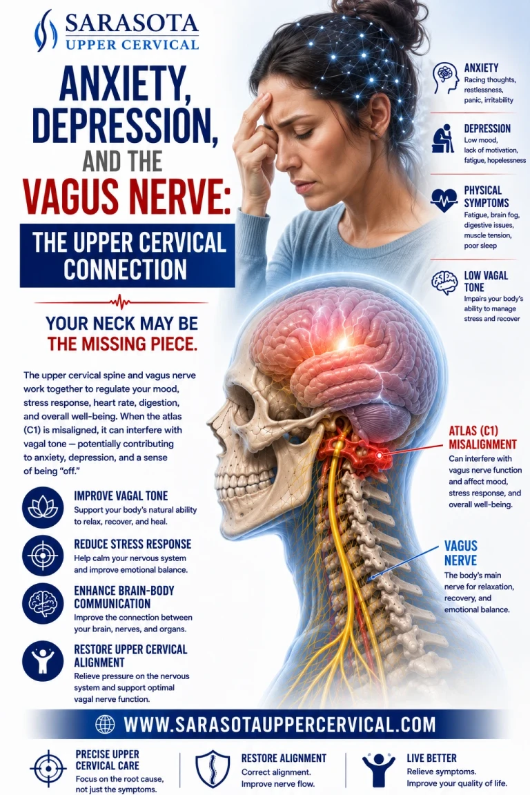
Anxiety and depression are almost always framed as chemical or psychological problems — a serotonin imbalance, a stressful life event, a pattern of negative thinking. What rarely gets discussed is the physical infrastructure underneath all of it: the vagus nerve, and the narrow corridor of the upper neck it must pass through on its way between the brainstem and the rest of the body. If that corridor is mechanically compromised, the nerve carrying most of the signal traffic between gut, heart, lungs, and brain can misfire — and mood is one of the first places that shows up.
The Anatomy: Where the Vagus Nerve Actually Lives
The vagus nerve (cranial nerve X) is the longest cranial nerve in the body, and unlike the other eleven, it doesn't stay in the head. It originates from three nuclei in the medulla oblongata — the lowest part of the brainstem. The dorsal motor nucleus of the vagus (DMNX) controls parasympathetic output to the heart, lungs, and digestive organs. The nucleus ambiguus controls the muscles of the throat, larynx, and soft palate, and also sends parasympathetic fibers to the heart. And the nucleus tractus solitarius (NTS) receives almost all of the sensory (afferent) information coming back up the vagus nerve from the body, relaying it directly into brain regions that regulate mood: the amygdala, hypothalamus, and locus coeruleus.
Schedule Your Appointment
Schedule appointmentThis last point matters enormously for anxiety and depression. About 80–90% of vagus nerve fibers are sensory, not motor — meaning the vagus nerve spends most of its bandwidth sending information up from the organs to the brain, not the other way around. The NTS is the first relay station for that incoming data, and it sits at the very base of the skull, directly adjacent to the same structures involved in upper cervical alignment.
After leaving the brainstem, the vagus nerve exits the skull through the jugular foramen, a small opening at the base of the occiput, shared with the internal jugular vein and the glossopharyngeal and accessory nerves. From there, it descends through the neck inside the carotid sheath — a fascial tube that also contains the internal carotid artery and internal jugular vein — running directly past the lateral masses of the atlas (C1) and the body of the axis (C2).
Related article
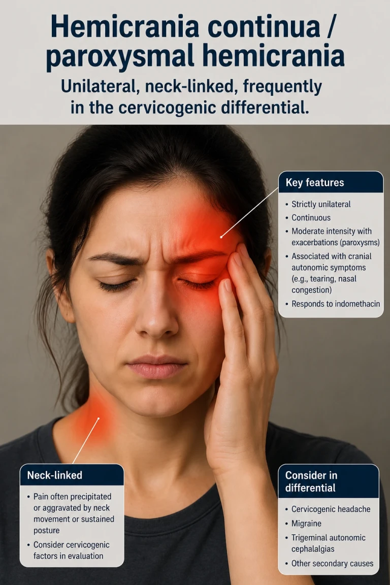
Is It a Cervicogenic Headache or Hemicrania Continua? How to Tell the Difference
Jun 13, 2026This is the anatomical detail that gets skipped in almost every mainstream discussion of the vagus nerve: for the first several inches of its journey, the vagus nerve is sitting in extremely close proximity to the upper two cervical vertebrae, wrapped in fascia that also attaches to the suboccipital muscles, the rectus capitis muscles, and the atlanto-occipital and atlanto-axial joint capsules.
Why the Upper Cervical Spine Matters
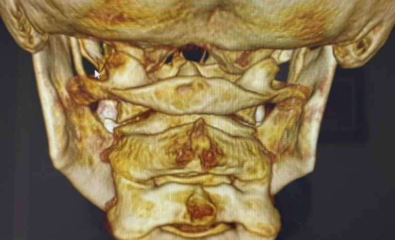
C1 and C2 are structurally different from every other vertebra in the spine. They have no intervertebral disc between them, they allow roughly 50% of all neck rotation, and they sit directly beneath the jugular foramen and the brainstem itself. Because of this anatomical relationship, a misalignment of C1 or C2 — even a subtle one, often invisible on standard imaging — can create fascial tension along the carotid sheath, subtly tensioning or compressing the vagus nerve as it runs alongside the jugular vein. It can restrict venous drainage through the jugular vein, which shares the same foramen and sheath as the vagus nerve, potentially affecting pressure dynamics around the brainstem. It can irritate the suboccipital muscles and upper cervical joint capsules, which are densely innervated and feed directly into the same brainstem regions (including the NTS) that process vagal sensory input. And it can alter proprioceptive input from the upper cervical spine, which has a disproportionately high density of mechanoreceptors compared to the rest of the spine and communicates directly with brainstem centers that regulate autonomic tone.
None of this requires a herniated disc or a dramatic injury. Upper cervical misalignment is frequently the result of whiplash, a fall, poor posture sustained over years, or a birth injury that was never addressed — and it can persist for decades without being detected by conventional X-rays or MRI, which are typically read for disc pathology, not for the millimeter-level misalignments that upper cervical specific care is designed to identify and correct.
Brainstem Irritation, Sympathetic Tone, and the Anxiety Pathway
The connection between the upper cervical spine and anxiety isn't limited to vagal afferent signal quality — it also runs through a second brainstem circuit that directly controls how much sympathetic ("fight or flight") drive the body is running on at any given moment: the rostral ventrolateral medulla (RVLM).
The RVLM is the primary sympathetic premotor center in the brainstem. It sends the signals that ultimately determine heart rate, vascular tone, and overall sympathetic outflow to the rest of the body. Critically, the RVLM does not operate independently — it sits under constant inhibitory control from the NTS, relayed through the caudal ventrolateral medulla (CVLM). Under normal conditions, this NTS → CVLM → RVLM pathway keeps sympathetic outflow in check: healthy afferent input into the NTS increases inhibitory drive onto the RVLM, holding sympathetic tone at an appropriate baseline. When that inhibitory input to the NTS is reduced or degraded, the brake on the RVLM eases, and sympathetic outflow increases — a mechanism well documented in baroreflex physiology, where a drop in inhibitory signaling to the NTS reliably produces a rise in sympathetic nerve activity.
This is the mechanical link that matters for anxiety. The NTS is the same nucleus that receives the vagus nerve's sensory input as it enters the brainstem — the same vagus nerve whose signal quality can be affected by fascial tension, venous congestion, or mechanical irritation at the jugular foramen and carotid sheath when C1 or C2 is misaligned. If irritation at this level of the brainstem and upper cervical spine degrades the quality or volume of inhibitory input reaching the NTS, the downstream effect is a chronic disinhibition of the RVLM — meaning the body's sympathetic output setting gets nudged upward and tends to stay there, independent of whatever is actually happening in the person's life or environment.
This matters because anxiety is not purely a top-down, cognitive phenomenon — a body of research on interoception and peripheral physiological feedback (going back to the James-Lange model and reflected in more recent "anxiety sensitivity" research) shows that the brain continuously monitors bodily arousal signals — heart rate, breathing rate, muscle tension — and uses them as raw material for the subjective experience of anxiety. When baseline sympathetic tone is chronically elevated because of brainstem-level disinhibition, the body is, in effect, constantly feeding the brain a stream of "threat-adjacent" physiological signals: elevated heart rate, shallow breathing, muscular readiness. The brain doesn't need a specific trigger to interpret that pattern as anxiety — the physiological substrate for anxiety is already present and elevated, which lowers the threshold at which any thought, situation, or stimulus gets tagged as threatening.
Put simply: when sympathetic tone is chronically elevated, anxiety becomes not just more likely but, in a sense, more physiologically primed — the nervous system is already sitting closer to the threshold where an anxious response gets triggered, before any psychological factor even enters the picture. This is one reason anxiety driven substantially by this pathway often doesn't respond fully to cognitive or behavioral intervention alone — those approaches work on the interpretation of the signal, not on the elevated sympathetic signal itself, if its origin is a mechanical, brainstem-level disinhibition rather than a purely cognitive or emotional one.
Connecting the Anatomy to Anxiety and Depression
Vagal tone — a measure of how well the vagus nerve is regulating the balance between the sympathetic ("fight or flight") and parasympathetic ("rest and digest") nervous systems — is consistently found to be lower in people with anxiety and depression. Low vagal tone is associated with reduced heart rate variability (HRV), a key biomarker of resilience to stress, and an impaired ability to downshift out of a stress response once a stressor has passed. It's also linked to increased circulating inflammatory markers, since the vagus nerve's cholinergic anti-inflammatory pathway is one of the body's primary brakes on systemic inflammation, and to disrupted gut-brain signaling, since the majority of serotonin production and gut microbiome communication with the brain travels via vagal afferents through the NTS.
If the vagus nerve's signal is being distorted at the point where it passes the upper cervical spine — before that signal ever reaches the NTS — the entire downstream chain of mood regulation is working with degraded information. This is the mechanical piece that's almost never addressed in a typical anxiety or depression treatment plan, which tends to focus entirely on neurotransmitter chemistry or cognitive patterns, both of which are real and important, but incomplete if there's an unresolved structural component upstream.
What This Means in Practice
This isn't a claim that upper cervical misalignment causes anxiety or depression outright, or that correcting it replaces therapy, medication, or lifestyle intervention where those are appropriate. It's a missing piece: for some people, particularly those with a history of neck trauma, whiplash, or postural strain who haven't responded fully to standard mental health treatment, an unaddressed upper cervical misalignment may be quietly limiting the nervous system's capacity to regulate itself — no matter how much work is being done elsewhere.
Upper cervical specific chiropractic care is focused entirely on identifying and correcting misalignment at C1 and C2, using precision imaging to measure exactly how the vertebrae are positioned relative to the skull and the rest of the spine — not generalized neck adjustment, but a specific, measured correction aimed at restoring proper alignment at the exact junction where the vagus nerve, the brainstem, and the upper cervical spine all meet.
If you've dealt with anxiety, depression, or mood symptoms that haven't fully responded to conventional care — especially alongside neck pain, headaches, or a history of trauma to the head or neck — it may be worth having your upper cervical alignment evaluated.


Leave a comment