

Serving Sarasota, Bradenton, and Lakewood Ranch
Two people describe an itch that will not stop. One points to the outside of both forearms. The other reaches over their shoulder to a spot beside the shoulder blade they cannot quite scratch. Neither has a rash. Neither responds to antihistamines. Both have been told, in one form or another, to moisturize and manage their stress.
Schedule Your Appointment
Schedule appointmentThese are two different conditions with two different names, two different nerve territories, and two different dermatomes. They are also, at the level that matters most, the same problem: a sensation manufactured somewhere along a spinal sensory pathway and then projected outward onto skin that has nothing wrong with it. Understanding how they differ is what allows correct identification. Understanding what they share is what explains why the spine keeps appearing in the literature for both.
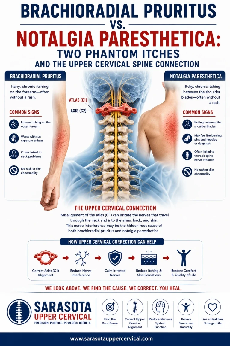
What brachioradial pruritus looks like
Brachioradial pruritus produces itching, burning, stinging, or tingling on the dorsolateral upper extremity — the outer forearm, sometimes extending to the upper arm and shoulder. It is bilateral in roughly three quarters of cases and most commonly maps to the C5 and C6 dermatomes, with involvement documented as far as C8. There are no primary skin lesions. The affected skin looks ordinary, and whatever marks appear are excoriations the patient created.
Its signature feature is the response to cold. Applying an ice pack produces immediate but temporary relief, a response distinctive enough that it functions as a clinical fingerprint separating this condition from other causes of itching. Its other defining feature is seasonality. Symptoms flare in summer and after ultraviolet exposure and often remit entirely in winter, which is why the condition was originally described in Florida in 1968 as a solar pruritus. It disproportionately affects fair-skinned, middle-aged women — with an approximate three-to-one female predominance and typical onset between forty-five and sixty-five — and it clusters among people with outdoor lifestyles: cyclists, boaters, golfers, gardeners, anyone whose forearms have accumulated decades of Gulf Coast sun.
Skin biopsy, when performed, tends to show reduced density of epidermal and dermal nerve fibers alongside actinic elastosis. The nerve supply to the itching skin is depleted rather than inflamed.
What notalgia paresthetica looks like

Notalgia paresthetica produces localized chronic itching medial or inferior to the scapula — the interscapular and infrascapular region of the upper and middle back. It is attributed to alteration of the cutaneous branches of the posterior rami, particularly the upper branches of the T2 through T6 spinal nerves. Alongside itching, patients report burning, coldness, numbness, tingling, allodynia, hyperalgesia, and hypoesthesia, which is the full vocabulary of a sensory neuropathy rather than a skin complaint.
Where brachioradial pruritus is usually bilateral, notalgia paresthetica is usually unilateral and, curiously, more often left-sided. It affects women predominantly, most commonly between the ages of fifty-four and sixty-two.
Its most visually distinctive feature is a well-circumscribed hyperpigmented patch in the affected area — the finding that most often brings patients to a dermatologist in the first place. This is worth understanding correctly, because it is frequently misread. There are no primary cutaneous lesions in notalgia paresthetica. The pigmentation is secondary to chronic scratching and rubbing of itchy, partly desensate skin over months and years. The mark is a record of the itching, not its cause.
One proposed mechanism specific to this condition involves the local anatomy of the back. The cutaneous branches of the thoracic dorsal rami penetrate the paraspinal musculature at close to a right angle on their way to the skin, passing through the multifidus and the muscles stabilizing the scapula. That geometry makes them mechanically vulnerable, and muscle strain, spasm, or postural loading in that region is thought to be capable of impinging them.
Telling them apart
The distinctions are practical and mostly reduce to location, laterality, appearance, and triggers.
If the itching is on the outer forearms, affects both sides, leaves no pigment change, flares in summer, worsens after a day outdoors, and calms within seconds under an ice pack, the picture favors brachioradial pruritus. If the itching sits beside or below one shoulder blade, is confined to one side, has produced a brown patch on otherwise normal skin, and shows no clear relationship to sun exposure or season, the picture favors notalgia paresthetica. Both are neuropathic, meaning both will fail antihistamines, both will look unremarkable on a routine skin exam apart from self-inflicted damage, and both are frequently missed for years.
There is no rule preventing a person from having both, and in patients with multi-level degenerative change, coexistence is entirely plausible.
Related article
Arm Numbness & Tingling Treatment in Sarasota, Bradenton, and Lakewood Ranch | Arm Pain Relief
Mar 06, 2026What they share, and why it points to the spine
Strip away the anatomy and both conditions are the same phenomenon. Neither itch originates in the skin. Both are generated by dysfunction in a sensory pathway and then referred outward to the dermatome that pathway serves, in the same way phantom limb sensation is referred to a hand that is no longer there. The brain has no mechanism for verifying where along a sensory line a signal was introduced. Activity in a C6 pathway is experienced as forearm itching whether it began at the wrist or in the dorsal horn.
The relay itself is shared. Pruriceptive afferents from either territory enter the dorsal horn, where second-order neurons release gastrin-releasing peptide onto gastrin-releasing peptide receptor neurons, which cross the midline and ascend in the spinothalamic tract toward the thalamus and on to the sensory cortex and insula. This relay is governed by inhibitory control from local interneurons and, importantly, by descending projections from the brainstem that directly suppress spinal gastrin-releasing peptide receptor neurons as part of an active anti-itch pathway. Itch is not simply transmitted upward — it is permitted upward. A great deal of low-grade peripheral noise is filtered out before it ever reaches awareness, and chronic itch can therefore arise either from excessive input at the periphery or from failure of suppression from above.
That shared architecture is the reason the same interventions appear in both literatures — gabapentinoids, tricyclics, topical capsaicin, cold — and the reason neither responds to anything aimed at the skin.
The spinal association is also shared, and it is better documented than most patients are told. In brachioradial pruritus, cervical involvement was first proposed in 1983 with the description of cervical vertebral osteoarthritis in affected patients, and subsequent imaging has repeatedly identified degenerative disc disease and foraminal stenosis capable of compressing the C5 through C8 roots. Case reports document complete resolution following cervical nerve root blocks at the levels of greatest stenosis and following surgical decompression in patients who had failed every drug. In notalgia paresthetica, degenerative cervical changes are considered associated with pathogenesis, and one comparative study found significantly more cervical degenerative change and more herniation at the C6–C7 segment in patients with the condition than in controls. Some authors have gone as far as to describe notalgia paresthetica as potentially a cutaneous sign of underlying degenerative cervical spine disease, and have proposed that first-line management for cases with associated cervical involvement may reasonably include spinal manipulation, physical therapy, massage, cervical traction, and cervical muscle strengthening before more invasive options.
An honest caveat that most articles omit
Here is the finding that any serious discussion of this topic has to include: degenerative changes on cervical imaging are extraordinarily common in people with no symptoms at all. One frequently cited figure places degenerative cervical findings in around ninety percent of asymptomatic subjects, alongside thoracic degenerative changes in roughly forty-seven percent. Critics of the radiculopathy model for notalgia paresthetica also point out, fairly, that a true radiculopathy would be expected to produce symptoms across a whole dermatome rather than the isolated patch that is typically seen.
This means that finding degeneration on a scan in someone with a neuropathic itch proves very little on its own. Almost everyone past middle age has some. The clinical question is never whether degeneration exists but whether the level and side of the finding correspond meaningfully to the level and side of the symptom, and whether the mechanical picture is plausible enough to be worth addressing. A practitioner who shows you a scan and declares the arthritis to be the cause of your itch is skipping a step that matters.
Where the upper cervical spine fits
Most of the published attention in both conditions falls on the lower cervical and upper thoracic segments, which is appropriate, because that is where the relevant roots exit and where degenerative narrowing concentrates. But that focus leaves two questions unaddressed, and both of them lead upward.
The first is mechanical. The atlas and axis establish the reference position of the head, and the segments beneath them organize themselves around whatever that reference happens to be. When upper cervical alignment is disturbed — after whiplash, a fall, a sports collision, or a slow postural drift — the lower cervical and cervicothoracic segments are drawn into compensation and carry load in a configuration they were not designed for. Sustained over decades, this is a coherent contributor to precisely the degenerative and foraminal changes that both literatures implicate. It offers an explanation for why one person's neck degenerates asymmetrically at C5–C6 or C6–C7 while another's does not.
The second is neurological, and it is the more interesting of the two. The upper cervical spine sits at the junction where the spinal cord becomes the brainstem, immediately adjacent to the structures generating the descending inhibitory control that gates the spinal itch relay. The suboccipital musculature spanning this region carries an exceptionally high density of proprioceptive receptors and delivers a continuous, high-resolution stream of positional information into brainstem sensory processing. Degraded or asymmetric input from a misaligned upper cervical segment offers a plausible route by which that descending suppression could become less effective — allowing peripheral signals that should have been filtered into silence to arrive in consciousness as an intractable itch.
This is a mechanistic hypothesis grounded in established neuroanatomy, not a demonstrated causal chain, and it deserves to be labeled as such. There are no established treatment guidelines for brachioradial pruritus, the cervical intervention evidence in both conditions consists largely of case reports and small series rather than controlled trials, and the chiropractic-specific literature on neuropathic itch is thin. What can be said with confidence is that these are neurological conditions, that spinal involvement is well documented in both, and that the neck is a region the standard dermatologic workup does not examine.
What a useful evaluation involves
Whichever of the two conditions is in play, the assessment follows the same logic. It begins with mapping the itching against neuroanatomical territory rather than accepting a general report — establishing whether the pattern is dermatomal, whether it is unilateral or bilateral, whether pigment change is present, whether cold relieves it, and whether it tracks with season and sun exposure. It moves to a detailed history of neck and upper back trauma, including events long since dismissed as unrelated, because no one previously asked about a car accident in the context of an itch. It includes direct examination of upper cervical alignment and segmental motion, with cone beam computed tomography where appropriate to assess the atlas and axis relationship in three dimensions rather than inferring it from a flat image. And it includes a frank assessment of whether the findings correspond to the symptom well enough to justify care, because sometimes they will not, and the right response in that case is a referral rather than a treatment plan.
If you have spent months or years being told that skin which looks completely normal is somehow the source of relentless itching, the more useful question is not what is wrong with the skin. It is what is happening to the nerve that serves it, and where along that nerve's path the trouble begins.
This article is for educational purposes only and does not constitute medical advice, diagnosis, or treatment. Chronic itching can be a symptom of systemic conditions including thyroid disease, liver and kidney disorders, iron deficiency, and certain blood disorders and malignancies, and warrants evaluation by a qualified healthcare provider. Individual results vary, and no specific outcome is promised or implied. Seek prompt medical attention for progressive weakness, numbness, loss of coordination, or changes in bowel or bladder function.
Dr. Drew Hall



Leave a comment