

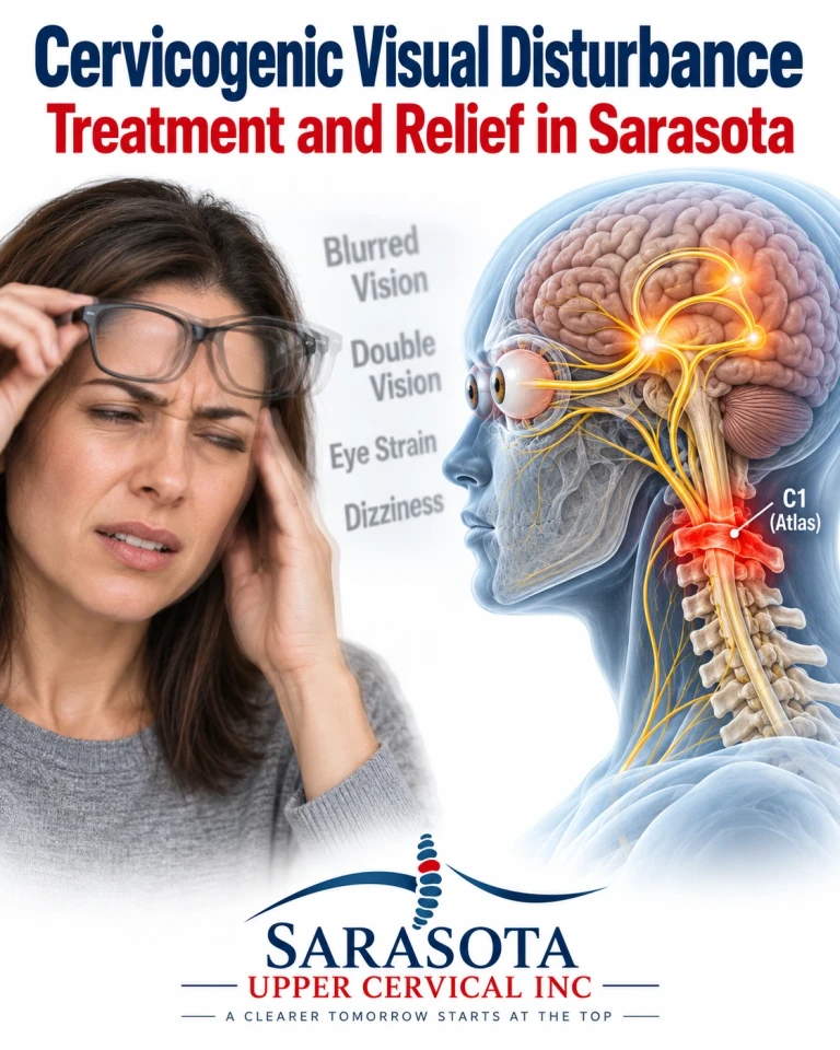
Blurred vision with neck movement is one of the more disorienting symptoms people bring to an upper cervical practice in Sarasota, Bradenton, and Lakewood Ranch, and one of the most frequently dismissed. The eye examination is normal. The prescription is correct. The retina is healthy. And yet vision goes soft when the head turns, text swims when reading, screens become exhausting, and the visual world feels subtly unreliable.
When the eyes are healthy but vision is not behaving, the problem is usually not in the eye. It is in the system that tells the eyes where the head is.
Schedule Your Appointment
Schedule appointmentWhy the neck is part of the visual system
Stable vision is not passive. To keep an image fixed on the fovea while the body moves, the brain must continuously calculate where the eyes are pointing relative to the world — which requires knowing where the head is on the neck, and where the neck is on the trunk. Three streams supply that calculation: vestibular input from the inner ear, visual input from the retina, and proprioceptive input from the body, most densely from the cervical spine.
These converge in the vestibular nuclei in the floor of the fourth ventricle, immediately above the craniocervical junction, and from there into the oculomotor nuclei that drive the eye muscles.
Two reflexes make the connection explicit. The vestibulo-ocular reflex generates compensatory eye movement from inner ear input. The cervico-ocular reflex generates compensatory eye movement from neck proprioception. They work together, and the cervical contribution is not a minor adjunct — the vestibular apparatus detects head movement in space but cannot distinguish head-on-neck rotation from whole-body rotation. Only cervical proprioception resolves that ambiguity.
The upper cervical segments carry most of this signal. The suboccipital muscles contain one of the highest muscle spindle densities found in the human body, and the atlanto-occipital and atlanto-axial joint capsules are richly innervated by C1 and C2 afferents. These structures are position sensors more than movers. An estimate of that spindle density comes substantially from anatomical specimen studies including fetal material, and I flag that because the figure is widely quoted without the caveat.
The test that demonstrates it
Related article
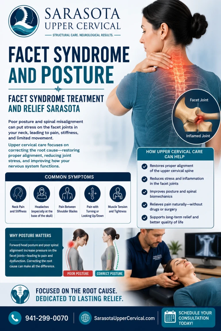
Facet syndrome and posture | Facet syndrome treatment and relief Sarasota
Jun 24, 2026
This is not only theory, and there is an objective measure.
The smooth pursuit neck torsion test compares eye-tracking performance in two positions. First the patient follows a moving target with head and trunk aligned. Then the trunk is rotated beneath a stationary head, so the vestibular and visual inputs are unchanged while cervical afferent input is altered. In a normal system, tracking performance barely differs between the two, because neck proprioception is not distorting the calculation.
Julia Treleaven and colleagues at the University of Queensland tested this in 100 people with persistent whiplash — 50 reporting dizziness and 50 not — against 50 healthy controls. Tracking difference between positions averaged 0.11 in the whiplash group with dizziness, 0.07 in the whiplash group without dizziness, and 0.01 in controls, with the differences statistically significant. The results were not explained by anxiety levels, though the authors noted both nociceptive and proprioceptive factors may contribute.
Two things are worth drawing out. The abnormality appeared even in whiplash patients who did not report dizziness — meaning cervically driven oculomotor disturbance can be present without the patient recognising it as a balance problem. And the mechanism the literature proposes is a mismatch between cervical proprioceptive input and its interconnections with the vestibular and visual systems.
An honest qualification belongs here. Findings across studies have not been uniform: some research groups have reproduced these differences and others have not, and reviewers attribute the inconsistency partly to a lack of methodological consensus on equipment and protocol. Reliability work suggests the test performs best at larger target amplitudes and slower velocities. It is a useful clinical measure with genuine supporting evidence, not a settled gold standard.
What patients actually describe
The symptom vocabulary is consistent once you know to listen for it. Vision that blurs or softens during head movement rather than at rest. Difficulty tracking a line of text, with words seeming to shift or requiring re-reading. Visual fatigue disproportionate to screen time. Discomfort in visually busy environments — supermarket aisles, patterned floors, traffic, scrolling. A sense that the visual world lags slightly behind head movement. Light sensitivity. Difficulty judging distance or catching objects.
Frequently these accompany neck pain, suboccipital tightness, restricted rotation, headache, or dizziness, and frequently they began after a whiplash injury or head impact.
Related article
What this is not
Several conditions produce visual disturbance and are not cervicogenic, and they need excluding first.
Refractive error and presbyopia are the obvious ones and are why an optometric examination comes before anything here. Dry eye produces fluctuating blur that improves with blinking. Cataract produces glare and gradual clouding. Convergence insufficiency produces blur and doubling at near, and is common after concussion. Vestibular disorders produce oscillopsia — the visual field appearing to bounce — which is a different symptom from blur on movement. Superior canal dehiscence produces visual disturbance triggered by loud sound or straining rather than by head position. Migraine produces visual aura with a characteristic build and duration. Optic neuritis produces painful visual loss and requires urgent assessment.
If your visual symptoms are triggered by sound, or occur at rest without any movement, or involve loss of vision rather than instability of vision, the cervical spine is not the explanation.
Stating the claim honestly
Cervical afferents contribute to oculomotor control. That is established, and the smooth pursuit neck torsion test provides an objective measure of disturbance in that contribution. Whiplash and other cervical injuries can produce measurable eye movement abnormalities.
What is less established is treatment. The literature on managing cervical sensorimotor disturbance comes largely from physiotherapy, using manual therapy combined with oculomotor and proprioceptive retraining, and the trials are small. There is no randomised trial of upper cervical chiropractic for cervicogenic visual disturbance. The reasoning that restoring upper cervical alignment improves the quality of proprioceptive input into the vestibular and oculomotor systems is coherent and anatomically grounded, and I am labelling it as reasoned rather than demonstrated.
What I would add is that retraining matters alongside any structural work. Gaze stability exercises, smooth pursuit training, and joint position sense retraining have supporting evidence in the cervical sensorimotor literature, and a passive approach alone is unlikely to be sufficient.
Related article
Precision and knowing when not to adjust
Upper cervical correction involves no twisting, no popping, no cracking, and no pulling of the neck. Cone beam computed tomography measures the three-dimensional position of the atlas and axis against your own anatomy so a correction is calculated rather than estimated.
Objective measures determine whether a correction is indicated and whether it is holding: leg length assessment, cervical range of motion, joint position error testing, balance measurement including tandem stance with eyes closed, and thermographic pattern tracking. When those show the correction is holding, no adjustment is given. In a system whose problem is distorted afferent input, adding unnecessary input is not neutral.
Red flags requiring urgent evaluation
Sudden loss of vision in one or both eyes, a curtain or shadow across the visual field, or a sudden increase in floaters with flashes of light requires emergency ophthalmological assessment. Painful eye movement with reduced vision suggests optic neuritis. Double vision that persists with one eye covered, or new double vision of sudden onset, requires urgent neurological assessment. Visual disturbance with severe headache, weakness, numbness, slurred speech, or facial droop requires emergency evaluation for stroke. Visual change with a red painful eye and haloes around lights may indicate acute glaucoma. Any visual symptom following significant head trauma warrants medical evaluation before manual care.
An evaluation in Sarasota
If your eye examination is normal and your vision still misbehaves when you move your head, particularly if this began after a whiplash injury or concussion, an upper cervical evaluation with sensorimotor testing may identify a contributor that the eye examination cannot detect. This works alongside your optometrist, ophthalmologist, and physician rather than in place of them, and an eye examination should come first. To arrange a consultation call 941 259-1891.
Dr. Drew Hall, Upper Cervical Chiropractor
Serving Sarasota, Bradenton, and Lakewood Ranch, Florida
This article is for general educational purposes only and is not medical advice, a diagnosis, or a treatment recommendation for any individual. Visual symptoms require examination by a qualified optometrist or ophthalmologist to exclude ocular and neurological causes before any other explanation is pursued. Upper cervical chiropractic is an area of focus within chiropractic; it is not a board-recognized specialty, and no claim of specialization or superiority is made or implied. No randomised trial evidence exists for upper cervical chiropractic in cervicogenic visual disturbance, and none is claimed here; the cervical contribution to oculomotor control is established physiology presented as reasoned rationale rather than demonstrated treatment mechanism, and findings on the smooth pursuit neck torsion test have not been uniform across research groups. Suboccipital muscle spindle density figures derive substantially from anatomical specimen studies including fetal material. Cone beam computed tomography is used as a precision measurement and analysis tool, not as a stand-alone diagnostic device, and does not assess visual function. Individual results vary and no specific outcome is guaranteed. Seek emergency care for sudden vision loss, a curtain across the visual field, flashes with new floaters, painful eye movement with reduced vision, new double vision, or visual change with headache, weakness, numbness, or slurred speech. Always consult a qualified healthcare provider regarding diagnosis and treatment.



Leave a comment