

Introduction
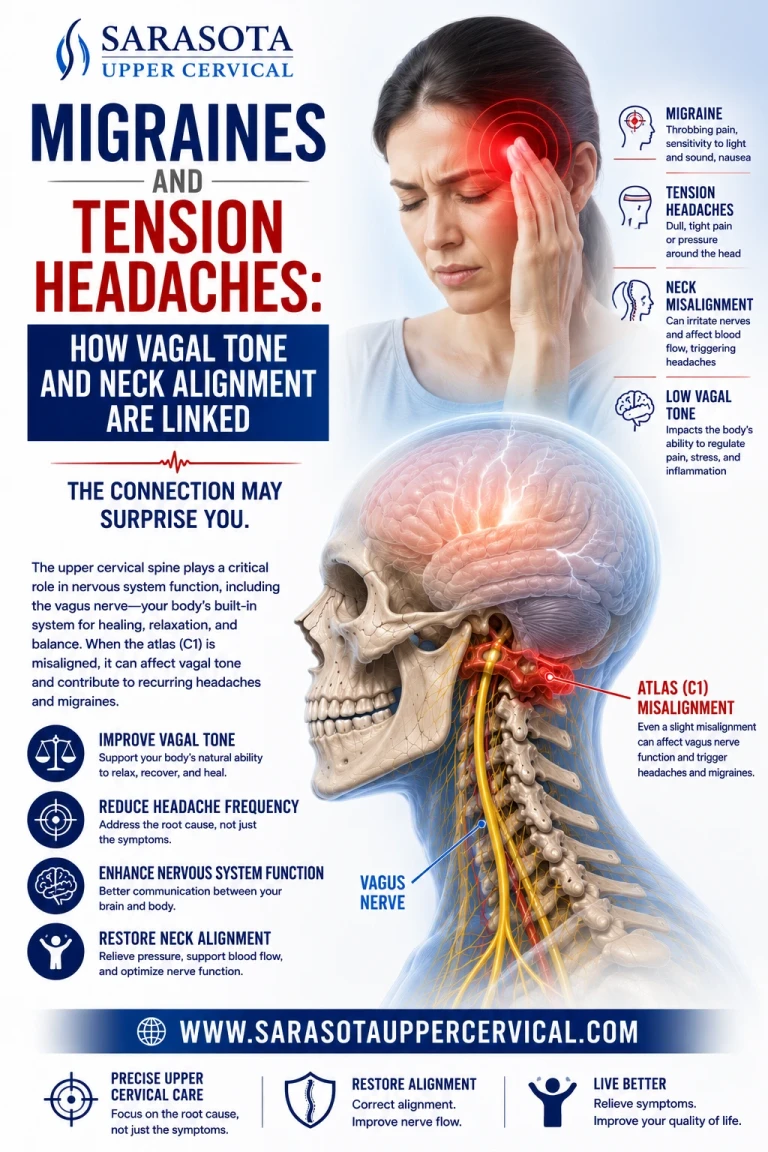
Migraine and chronic tension headache treatment tends to focus on triggers, medication, and pain management — rarely on the mechanical relationship between the upper neck and the brainstem structures that regulate both pain processing and autonomic (vagal) tone. Anyone who has ever noticed that their headaches worsen with poor posture, neck stiffness, or after whiplash has already brushed up against a connection that goes much deeper than "tight muscles."
The Anatomy: Trigeminocervical Convergence and the Vagus Nerve
To understand why headaches and the vagus nerve are connected at all, you have to understand a structure called the trigeminocervical nucleus. This is a region in the upper cervical spinal cord (spanning roughly C1 to C3) where sensory fibers from the trigeminal nerve (cranial nerve V, responsible for facial and head pain, including migraine pain) converge with sensory fibers from the upper cervical spinal nerves. Because these two systems physically converge on the same neurons, pain signals from the neck and pain signals from the head become functionally indistinguishable to the brain — this is why upper cervical dysfunction can generate headache and migraine-like pain, and why migraine can cause referred neck pain.
Schedule Your Appointment
Schedule appointmentThe vagus nerve sits immediately adjacent to this entire system. It exits the skull through the jugular foramen at the base of the occiput — a few centimeters from where the trigeminocervical nucleus is doing its convergence work — and descends through the neck within the carotid sheath, running directly past the atlas (C1) and axis (C2). The vagus nerve's sensory fibers terminate in the nucleus tractus solitarius (NTS), which sits in the same brainstem region as, and has established connections to, the trigeminocervical nucleus and the descending pain-modulation pathways that determine whether a headache signal gets amplified or dampened.
This is a critical anatomical detail: the brainstem doesn't process headache pain and vagal/autonomic tone in separate silos. They share overlapping neural real estate at the exact level of the upper cervical spine, which is why migraine is so often accompanied by autonomic symptoms, nausea, light sensitivity, hyperacusis, dizziness, cold extremities — that have nothing to do with pain perception directly, but everything to do with vagal and autonomic disruption happening in parallel.
Book a FREE consult by clicking here.
Why C1 and C2 Specifically
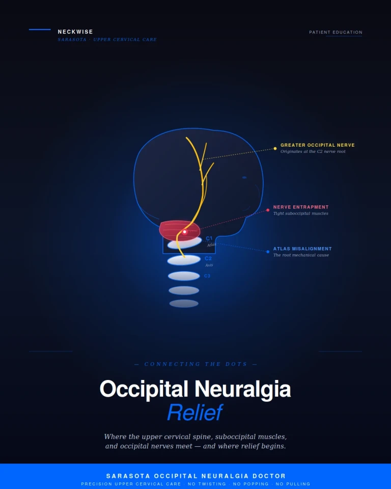
The atlas and axis are unlike any other vertebrae in the spine:
1.They have no intervertebral disc between them and account for roughly half of all cervical rotation
2.They sit in direct proximity to the jugular foramen, the vertebral arteries, and the vagus nerve as it exits the skull
3.The suboccipital muscles connecting the skull to C1 and C2 have one of the highest densities of muscle spindles (proprioceptors) anywhere in the body — far higher than muscles of similar size elsewhere in the spine
4.These proprioceptors feed directly into the trigeminocervical nucleus and brainstem centers involved in both pain gating and autonomic regulation
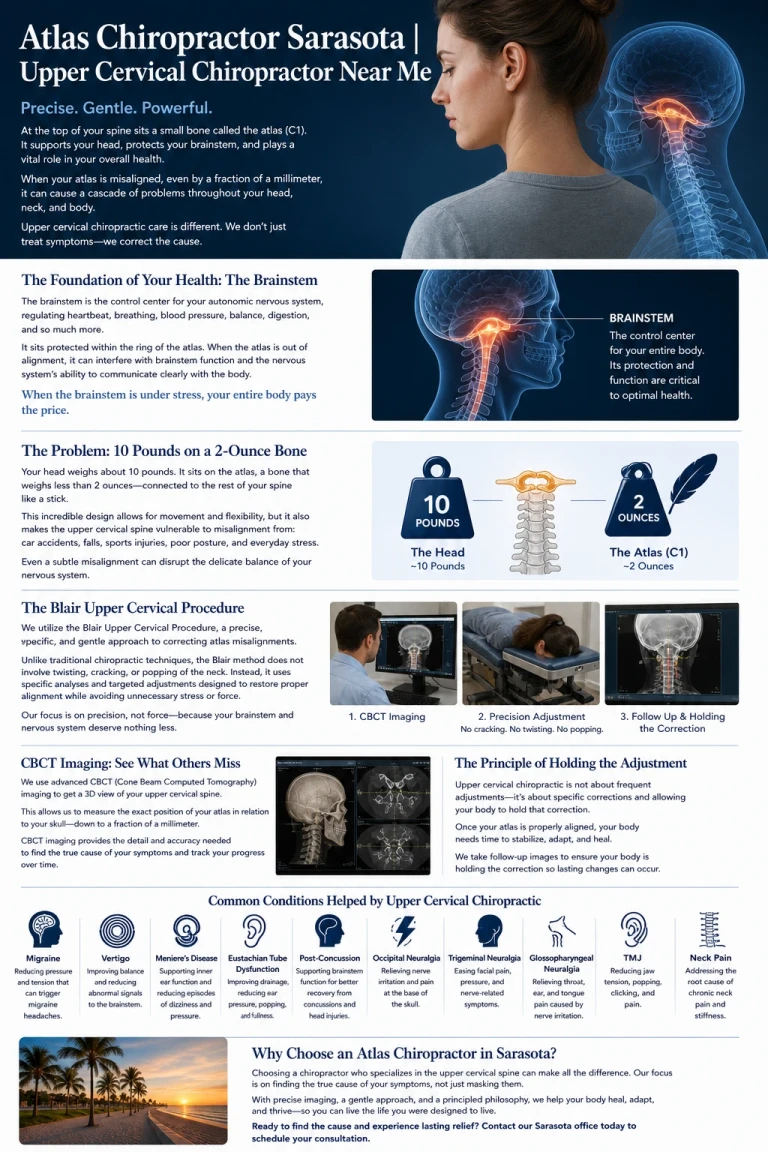
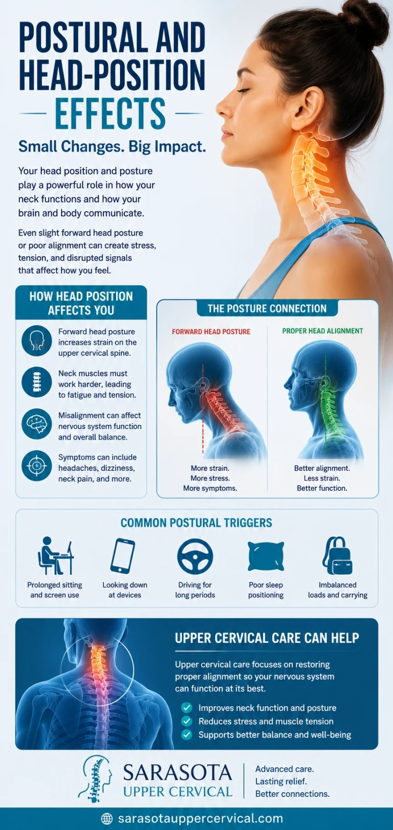
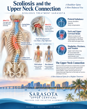
When C1 or C2 sits misaligned — even by a few millimeters, which is common after whiplash, a fall, or repetitive postural strain and frequently invisible on standard imaging — the effects cascade in two directions simultaneously:
1.Constant low-grade nociceptive (pain) input floods into the trigeminocervical nucleus from the misaligned joint and surrounding muscle tissue, lowering the threshold at which the brain interprets subsequent stimuli as head pain — a phenomenon called central sensitization
2.Mechanical tension along the carotid sheath and jugular foramen, where the vagus nerve travels, can alter the sensory signal quality reaching the NTS, disrupting the normal vagal brake on the sympathetic nervous system
3. The result is a nervous system primed for both pain amplification and poor autonomic regulation at the same time — which maps closely onto the clinical picture of chronic migraine: recurring head pain paired with nausea, dizziness, temperature dysregulation, and stress intolerance.
Vagal Tone's Direct Role in Migraine
Vagal tone, measured through heart rate variability (HRV), is consistently found to be reduced in people with chronic migraine compared to headache-free populations. Low vagal tone corresponds to:
1.Reduced capacity to down-regulate the sympathetic nervous system once a migraine attack begins, prolonging the episode
Elevated baseline inflammatory signaling, since the vagus nerve's cholinergic anti-inflammatory pathway is a primary regulator of systemic and neurogenic inflammation — and neurogenic inflammation around the trigeminal vasculature is a core mechanism in migraine itself
2. Disrupted gut motility and nausea, both hallmark migraine symptoms, since vagal efferents directly control gastric emptying
This is why vagus nerve stimulation devices have been explored as a migraine treatment with some clinical success — they attempt to externally restore a signal that, in many patients, may be getting interrupted mechanically before it ever needs external stimulation in the first place.
What This Means in Practice
Standard migraine treatment, medication, trigger avoidance, lifestyle modification, addresses real and important pieces of the picture, but rarely investigates whether an upper cervical misalignment is actively feeding both the pain-amplification pathway (via the trigeminocervical nucleus) and the autonomic-dysregulation pathway (via the vagus nerve and NTS) at the same time.
Upper cervical specific chiropractic care uses precision diagnostic imaging to measure the exact position of C1 and C2 relative to the skull and lower spine, and delivers a specific, measured correction — not a generalized neck adjustment — aimed at removing the mechanical interference at the exact junction where the trigeminocervical system, the vagus nerve, and the upper cervical spine converge.
If your migraines or tension headaches are accompanied by neck stiffness, a history of whiplash or head injury, or autonomic symptoms like nausea, dizziness, or temperature intolerance, it may be worth having your upper cervical alignment evaluated as part of the full picture.
book a FREE consult click here.


Leave a comment