

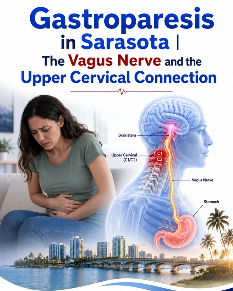
Gastroparesis is a serious motility disorder, and any discussion of it in a chiropractic context has to begin by saying so plainly. Delayed gastric emptying in the absence of mechanical obstruction produces nausea, vomiting, early satiety, bloating and epigastric pain, and in its more severe forms it causes malnutrition, dehydration, unstable blood glucose, and hospitalisation. It is managed by gastroenterology. What follows is an examination of one specific question — whether upper cervical structural dysfunction could plausibly contribute to vagal dysfunction in some patients — and an honest account of how much evidence stands behind that idea, which is considerably less than you will find claimed elsewhere.
What Gastroparesis Is and What Is Known to Cause It
Schedule Your Appointment
Schedule appointment
Gastric emptying depends on a coordinated system: the vagus nerve supplying extrinsic parasympathetic input, the enteric nervous system providing local control, the interstitial cells of Cajal generating the electrical slow waves that pace gastric contraction, nitrergic neurons mediating relaxation, and the smooth muscle itself executing the contraction. Gastroparesis is what happens when that system fails, and the literature is clear that failure can occur at several points.
The pathology is well characterised. Loss or depletion of interstitial cells of Cajal is a predominant finding, present in roughly half of cases and correlated with delayed emptying. Loss of nitric-oxide-producing neurons, vagal nerve impairment, changes in enteric neurons, and smooth muscle abnormalities have all been demonstrated. This is established pathology, drawn from tissue studies in human patients.
The recognised categories are diabetic, post-surgical, and idiopathic, with idiopathic accounting for the majority of cases. Diagnosis requires objective evidence of delayed emptying — gastric emptying scintigraphy remains the reference standard — after mechanical obstruction has been excluded by endoscopy or imaging.
The Vagus Nerve and Gastric Emptying
The vagal contribution to gastric function is not a matter of interpretation. Preganglionic parasympathetic fibers arise from the dorsal motor nucleus of the vagus in the medulla, descend through the neck, and synapse with enteric neurons in the stomach wall. Vagal afferents carry sensory information about distension and nutrient content back to the nucleus tractus solitarius. This circuit governs receptive relaxation, antral contraction, and pyloric coordination — the three things that have to work for a meal to leave the stomach on schedule.
The clearest demonstration that vagal integrity matters is surgical. Post-surgical gastroparesis is a recognised category precisely because operations that damage the vagus — historically vagotomy for ulcer disease, and more recently fundoplication and other upper gastrointestinal procedures — produce delayed gastric emptying as a known complication. Cut the vagus, and the stomach empties poorly. That is about as direct a causal demonstration as clinical medicine offers.
There is also interventional evidence in the other direction. An open-label pilot study of non-invasive vagal nerve stimulation in patients with idiopathic gastroparesis found improvement in cardinal symptoms and accelerated gastric emptying in a subset of patients. I want to be exact about what that study was, because it is misrepresented on at least one competing website in this area: it used transcutaneous electrical stimulation of the cervical vagus nerve. It was not a study of manual therapy, spinal adjustment, or chiropractic care of any kind, and it should not be cited as though it were. What it establishes is narrower but still relevant — that modulating vagal activity at the level of the neck can measurably change gastric emptying.
Where the Upper Cervical Spine Enters the Picture
The anatomical premise is not speculative. Both vagus nerves exit the skull through the jugular foramen and descend within the carotid sheath, running between the internal jugular vein and the carotid artery, immediately anterior to the upper cervical vertebrae. The transverse process of the atlas is close enough to this region that it serves as a surgical landmark during skull base procedures. The vagal ganglia — the superior (jugular) and inferior (nodose) — sit at the top of that descent, in the immediate vicinity of the atlas.
Related article
Piriformis Syndrome Treatment in Sarasota | Sciatica and the Upper Cervical Root Cause
Aug 13, 2026More usefully, there is published evidence that the atlas transverse process can mechanically compress a structure inside the carotid sheath. In a series of forty-six patients with cervical spondylosis-related internal jugular vein stenosis, just over half of the stenotic vessels were compressed by the C1 transverse process, and a further group by the C1 transverse process in combination with the styloid process. A separate surgical case series found that resecting the C1 transverse process restored venous outflow and relieved symptoms.
That is a meaningful finding for this discussion, and it is worth being precise about why. It establishes that the atlas transverse process is capable of exerting compressive force on carotid sheath contents. The vagus nerve travels in that same sheath. What it does not establish is that the vagus is similarly affected — the internal jugular vein is a thin-walled, compressible vessel, and a nerve is not. Documented vagal compression at this level comes chiefly from space-occupying lesions such as paragangliomas and schwannomas, not from vertebral position.
The Traction Hypothesis
The specific proposition — that loss of cervical lordosis places the vagus nerve under sustained longitudinal tension where it passes the atlas — now has a named formulation in the peer-reviewed literature. Hauser, Matias and Rawlings published "Cervicovagopathy" in Frontiers in Neurology in July 2025, proposing that progressive stretching of the posterior cervical ligaments produces a conduction block in the vagus, progressing to ligamentous instability, breakdown of the cervical curve, and eventually vagal degeneration measurable as reduced nerve cross-sectional area on carotid sheath ultrasound. They present measurements from a retrospective chart review of 234 consecutive patients.
This is the closest thing to published support the traction mechanism has, and it deserves to be described accurately rather than oversold. It is explicitly framed by its own authors as a proposed etiology — the word "potential" appears in the title. The supporting data is a retrospective chart review without a control group, not a controlled trial. The authors practice at a clinic offering the treatments the paper recommends, which is a relevant interest to disclose. And gastroparesis specifically was not among the nine symptoms examined in that chart review.
So: the traction hypothesis is a published hypothesis with preliminary uncontrolled observational support, not a demonstrated mechanism. A randomised trial examining heart rate variability after cervical sagittal alignment restoration has been registered but had not begun recruiting at the time of writing, which is a fair indication of where this question actually sits.
The Sympathetic Hypothesis
There is a second route that does not depend on mechanical compression at all, and I find it the more defensible of the two.
Sympathetic outflow inhibits gastrointestinal motility. That is established autonomic physiology: sympathetic activation reduces gastric contraction and slows transit, which is why digestion shuts down under acute stress. If upper cervical dysfunction raises resting sympathetic tone, delayed gastric emptying follows without any need for the vagus to be physically stretched or compressed.
The relevant experimental work is directly on point. Bolton, Kerman, Woodring and Yates, publishing in Brain Research Bulletin in 1998, stimulated the C2 dorsal root ganglion and the C2 and C3 branches supplying dorsal neck muscles in anaesthetised cats, and recorded the response in the splanchnic sympathetic nerve — the sympathetic supply to the abdominal viscera, including the stomach. Upper cervical afferent input produced measurable changes in sympathetic outflow to the gut. The suboccipital region is well suited to generating that input, carrying muscle spindle densities that Kulkarni and colleagues measured between 98 and 242 per gram, against roughly 17 for opponens pollicis, though those figures came from fetal specimens.
Two caveats, stated because they matter. Bolton's data showed cervical input could either excite or inhibit splanchnic sympathetic activity depending on conditions — it is not a simple dial. And this was an anaesthetised animal preparation demonstrating an acute reflex, not a chronic clinical state. That a sustained upper cervical misalignment produces enough sympathetic elevation to meaningfully delay gastric emptying in humans is a reasoned hypothesis extrapolated from established physiology. It has not been tested.
The same framework underlies our discussion of the brainstem, sympathetic tone, and sleep, where the mechanism is described in fuller detail.
What the Evidence Does Not Show
There are no randomised controlled trials of upper cervical chiropractic care for gastroparesis. There are no controlled trials of any chiropractic intervention for gastroparesis. There is no published study demonstrating accelerated gastric emptying on scintigraphy following cervical adjustment, and any page claiming otherwise is either mistaken or has borrowed a result from the electrical stimulation literature.
Upper cervical care is therefore not a treatment for gastroparesis and should not be presented as one. What can be said honestly is narrower: gastroparesis is substantially a disorder of autonomic control of the stomach; the vagus nerve and sympathetic outflow to the viscera both pass through territory anatomically related to the upper cervical spine; and in a patient with documented craniocervical trauma, a demonstrated structural finding, and gastroparesis that has resisted standard management, investigating that structure is defensible. Whether correcting it changes anything is an individual question answered by measurement, not by a promise made in advance.
Objective Testing
Nothing above justifies treatment without measurement. We image the craniocervical junction with cone beam computed tomography, which reconstructs volumetric anatomy rather than requiring three-dimensional position to be inferred from a flat radiographic shadow. The honest qualifiers are that effective dose varies with field of view and protocol, and that CBCT images bone — it does not visualise the vagus nerve, and it cannot demonstrate that a nerve is under tension.
Heart rate variability provides an accessible measure of autonomic state, and it is the appropriate before-and-after metric here given that the entire hypothesis concerns autonomic balance. It should be recorded before care begins and tracked through a defined trial period. Gastric emptying, if it is going to be assessed at all, is assessed by scintigraphy ordered through your gastroenterologist — not by us, and not by inference from symptoms.
A principle worth stating: if a defined trial of care produces no measurable change, that is an answer, and the correct response is to say so rather than to extend the trial.
Correction Without Twisting, Popping, or Pulling
The correction involves no twisting of the neck, no forced rotation, no pulling or traction of the head, and no cracking or popping, because no joint cavitation is induced. The patient lies on their side while a low-force contact — measured in ounces rather than pounds — is applied to the atlas along a vector calculated from their own imaging.
Related article

Upper Cervical Chiropractor Sarasota, Bradenton, Venice and Tampa Bay Area
Nov 15, 2021A related principle is what we call holding the adjustment. The objective is not frequent repetition but stability: once the atlas holds its corrected position, no further correction is given. Patients are reassessed to determine whether the correction is holding, and adjusted only when it is not. This matters particularly for patients with significant ligamentous laxity or suspected instability, in whom repeated force is a hazard rather than a benefit, and high-velocity rotational manipulation is contraindicated outright.
When It Is Not the Neck, and When It Is an Emergency
This section carries more weight in gastroparesis than in most conditions, because the consequences of delay are real.
Gastroparesis is diagnosed and managed by gastroenterology. Mechanical gastric outlet obstruction must be excluded before any functional diagnosis is entertained. Diabetic gastroparesis requires glycaemic management, and the relationship runs both ways, since erratic emptying destabilises blood glucose. Medication-induced delayed emptying is common and frequently overlooked — opioids, GLP-1 receptor agonists, anticholinergics, and certain antidepressants all slow the stomach, and reviewing the medication list is often more productive than any structural investigation. Cyclic vomiting syndrome, cannabinoid hyperemesis, eating disorders, hypothyroidism, and connective tissue disorders including Ehlers-Danlos syndrome all belong on the differential.
Seek medical care urgently, not chiropractic care, for persistent vomiting preventing fluid intake, signs of dehydration, vomiting blood or material resembling coffee grounds, severe or worsening abdominal pain, unintentional weight loss, inability to keep down medication, or blood glucose you cannot control. Gastric bezoars and aspiration are recognised complications. And as with any craniocervical presentation, progressive neurological deficit, difficulty swallowing or speaking, or suspicion of craniocervical instability requires medical evaluation rather than adjustment.
Do not stop or alter prescribed treatment on the basis of anything in this article.
Evaluation in Sarasota, Bradenton, and Lakewood Ranch
If you are under gastroenterological care for gastroparesis, if your symptoms began or worsened after a car accident, fall, concussion or sports injury, and particularly if you have accompanying signs of autonomic dysregulation — orthostatic intolerance, temperature dysregulation, unexplained tachycardia, headaches originating at the skull base — then an upper cervical evaluation can tell you whether a structural finding exists. It is an additional line of investigation alongside your medical care, not a replacement for it. To learn more or to schedule call 941 259-1891.
Medical Disclaimer
This article is provided for general educational purposes only and does not constitute medical advice, diagnosis, or treatment, and does not establish a doctor-patient relationship. Gastroparesis is a serious medical condition requiring diagnosis and management by a qualified physician. Upper cervical chiropractic care is not a treatment or cure for gastroparesis or any other named disease, and no guarantee of any particular result is made or implied. Do not discontinue or alter any prescribed medication or treatment without consulting the prescribing physician. Individual results vary, and the mechanisms described in this article include both established physiology and hypotheses that have not been demonstrated in clinical trials, as explicitly indicated throughout the text. In accordance with Florida Administrative Code Rule 64B2-15.001, this content is offered without any claim of superiority of one method of treatment over another and without any promise of cure or guaranteed outcome.
Written by Dr. Drew Hall, upper cervical chiropractor, Sarasota Upper Cervical / Hall Upper Cervical Chiropractic PC, serving Sarasota, Bradenton, and Lakewood Ranch, Florida.


Leave a comment