

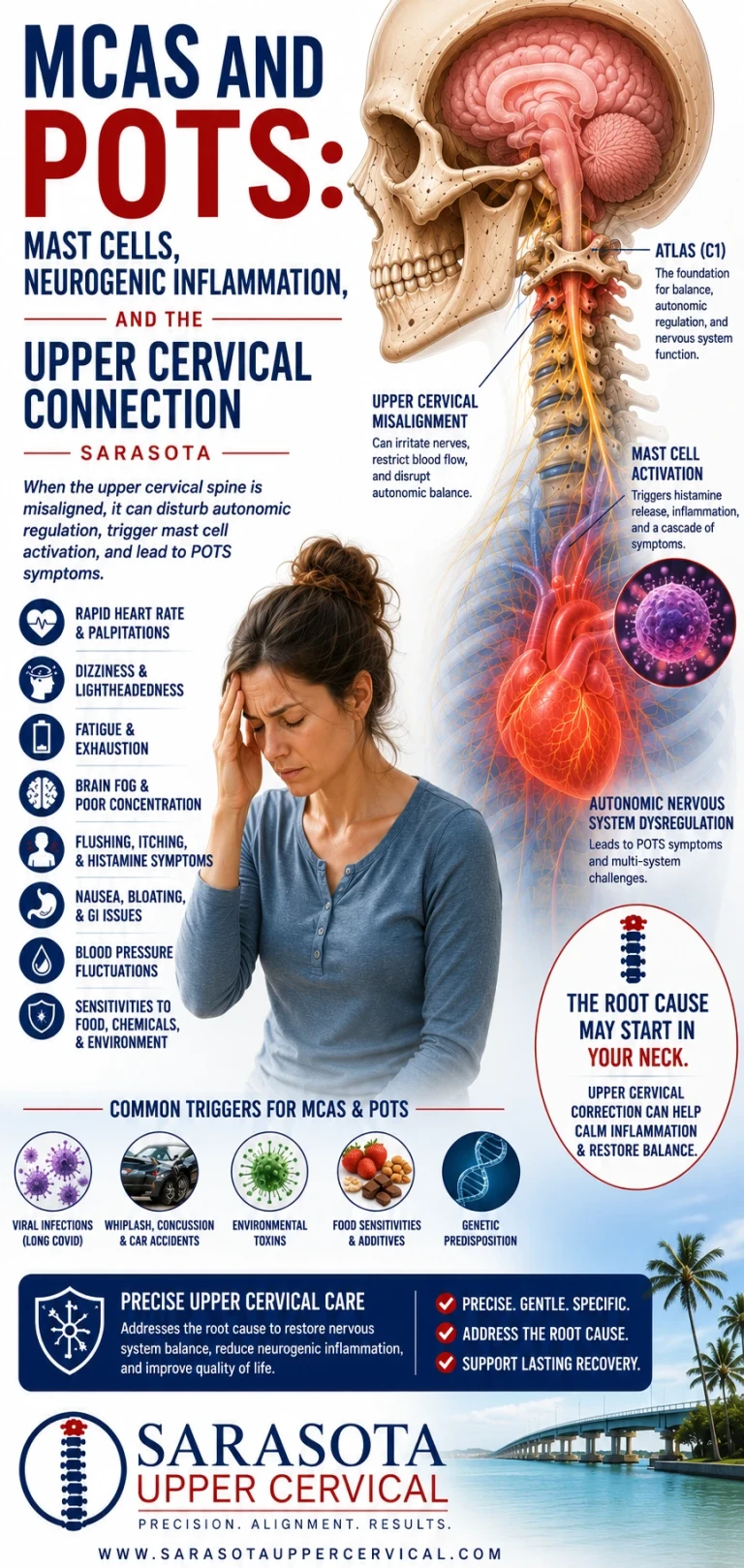
MCAS and POTS: Mast Cells, Neurogenic Inflammation, and the Upper Cervical Connection
Some people with POTS have a second layer to their illness that standard autonomic management does not touch. They flush — not blush, flush, in blotchy red waves across the chest, neck, and face. They react to foods that were fine last month. Heat, exercise, alcohol, fragrance, or stress sets off episodes involving racing heart, lightheadedness, nausea, abdominal cramping, diarrhea, and a sense of impending doom that lasts far longer than a panic attack. They may have hives, itching, or unexplained anaphylaxis-like reactions. Antihistamines help more than anyone expected them to.
Schedule Your Appointment
Schedule appointmentThat pattern points toward mast cell activation, and the overlap between mast cell activation syndrome and POTS is substantial enough that in some clinical populations it is closer to the rule than the exception.
This article explains what mast cells are doing, why they and the autonomic nervous system are so tightly coupled, what the research does and does not establish, and where the upper cervical spine fits into a picture that is fundamentally about the relationship between nerves and immune cells.
What mast cells are and why they matter here
Mast cells are tissue-resident immune cells packed with granules containing histamine, tryptase, heparin, prostaglandins, leukotrienes, and a long list of cytokines. They sit in the tissues that interface with the outside world — skin, gut lining, airways — and also in perivascular tissue throughout the body, positioned along blood vessels and, importantly for this discussion, alongside nerves.
When mast cells degranulate, they dump vasoactive mediators into local tissue and the circulation. Histamine dilates blood vessels and increases capillary permeability. Prostaglandins and leukotrienes amplify the effect. The immediate cardiovascular consequence of significant mast cell degranulation is vasodilation and fluid shifting out of the vasculature into tissue — which is to say, a sudden drop in effective circulating volume and vascular tone.
Now consider what that means in someone whose autonomic system is already struggling to defend blood pressure against gravity. A mast cell episode does not just cause flushing and itching. It actively undermines the exact physiological variables a person with POTS is fighting to maintain. And the body's response to sudden vasodilation is compensatory sympathetic activation — heart rate up, norepinephrine up, vasoconstrictive effort up.
This is why the two conditions amplify each other so effectively, and why treating one while ignoring the other so often fails.
What the research actually shows
Related article
Hypovolemic POTS: Low Blood Volume, Blood Pooling, and the Upper Cervical Connection | Sarasota
Jul 15, 2026
The foundational clinical work here is Shibao and colleagues in 2005, who evaluated 177 POTS patients and identified a subgroup with mast cell activation, defined as meeting POTS criteria plus having elevated urinary methylhistamine during flushing episodes. Eight female patients met criteria for both POTS and MCAS. A further sixteen had POTS and flushing without measurable methylhistamine elevation. In the group with confirmed elevation, both H1 and H2 histamine receptor antagonists were beneficial — and these patients also showed elevated norepinephrine levels, demonstrating a hyperadrenergic phenotype.
That last detail is the important one. Shibao's group suggested that mast cell activation could be producing the hyperadrenergic picture — that the increased norepinephrine was a response to mast-cell-driven vasodilation rather than a primary autonomic defect. The study also found that the primary problem in these patients was not diffuse autonomic neuropathy; their autonomic reflexes were intact or overactive.
That has a direct clinical implication. If you have been diagnosed with hyperadrenergic POTS and you flush, the possibility worth considering is that your sympathetic overdrive is downstream of mast cell activity rather than independent of it — which changes what interventions are likely to help.
Beyond that, the picture becomes murkier and the honest summary is that the association is real but poorly quantified. One retrospective review of 195 patients with both EDS and POTS reported that 31 percent also had MCAS compared with 2 percent of healthy controls — but the authors of the review discussing that finding noted explicitly that the criteria used for defining MCAS in that study were not clear. That caveat matters, because MCAS diagnostic criteria remain genuinely contested. Consensus criteria require episodic symptoms across multiple organ systems, objective evidence of mediator release such as elevated serum tryptase measured during an episode against baseline, and response to mast-cell-targeted therapy. Broader criteria used in some clinical settings produce dramatically higher prevalence estimates. When you encounter a striking statistic about MCAS prevalence in POTS, the first question to ask is which definition was used.
The proposed triad of POTS, MCAS, and hypermobile Ehlers-Danlos syndrome appears repeatedly in the literature, though it is described in reviews as arising from sporadic reports and anecdotal observation, with the common mechanism still unidentified. It is a pattern worth recognizing clinically. It is not yet a established syndrome with a known cause.
The nerve-mast cell relationship
Here is the part that connects this to the nervous system, and it is genuinely well documented.
Mast cells and nerves are not merely neighbors. They form direct membrane-to-membrane contacts in living tissue, described in the literature since the late 1980s. This is a physical, structural relationship — the immune cell and the neuron are in contact.
Sensory nerves release neuropeptides, principally substance P and calcitonin gene-related peptide. Mast cells express receptors for both. Substance P and CGRP are potent histamine releasers from mast cells, acting through mechanisms independent of the classical IgE pathway. Substance P acts on mast cells through the neurokinin-1 receptor and also through MRGPRX2, a receptor increasingly recognized as central to substance-P-mediated neurogenic inflammation.
The consequence is that nerve activity can trigger mast cell degranulation directly. This is the basis of neurogenic inflammation: a sensory nerve fires, releases neuropeptides at its terminal, and mast cells nearby respond by releasing histamine and other mediators. Those mediators then sensitize the same sensory nerves, lowering their firing threshold, which increases neuropeptide release, which activates more mast cells.
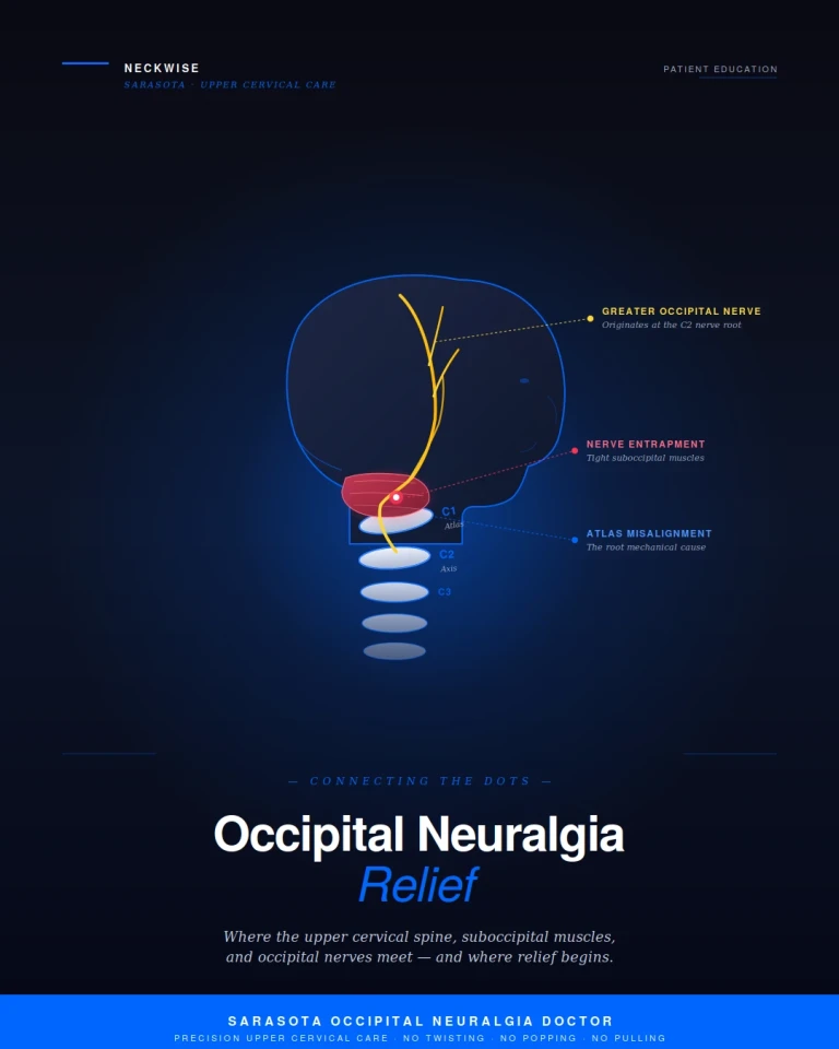
That is a positive feedback loop between the sensory nervous system and the immune system, and once established it is self-sustaining. It does not require an external allergen. Nerve traffic alone can drive it.
Traffic runs the other direction too. Mast cell mediators including histamine, cytokines, and proteases sensitize sensory neurons and contribute to neuroinflammation. And the autonomic nervous system modulates mast cell behavior — the vagus nerve is the efferent arm of the cholinergic anti-inflammatory reflex, in which vagal signaling suppresses pro-inflammatory mediator release from immune cells through alpha-7 nicotinic acetylcholine receptors. Reduced vagal tone means reduced restraint on inflammatory cells.
So the relationship is bidirectional and layered. Sympathetic overdrive and low vagal tone create conditions favorable to mast cell activation. Mast cell activation destabilizes blood pressure and provokes sympathetic compensation. Each drives the other.
Where the upper cervical spine enters
This is where careful language matters, because it would be easy to overstate.
There is no evidence that upper cervical misalignment causes mast cell activation syndrome. MCAS is an immunological disorder, and nothing about spinal alignment explains clonal mast cell populations, hereditary alpha-tryptasemia, or the genetic factors implicated in mast cell disorders. Any claim otherwise would be nonsense.
What can be said is narrower and rests on the documented relationship between autonomic tone and mast cell behavior.
The brainstem structures governing autonomic balance sit directly above the craniocervical junction. The nucleus tractus solitarius receives cardiovascular and visceral afferent information via the glossopharyngeal and vagus nerves and functions as the integrating hub for autonomic regulation. From the NTS, projections run to the caudal ventrolateral medulla, which inhibits the rostral ventrolateral medulla, the primary source of sympathetic outflow. Parasympathetic cardiac output originates in the nucleus ambiguus and dorsal motor nucleus of the vagus. Sympathetic and parasympathetic balance is set here, a few centimetres above the atlas.
Cervical afferents have documented access to this circuitry. Bolton and colleagues showed in 1998 that stimulating the C2 dorsal root ganglion and the C2 and C3 branches innervating the dorsal neck muscles produced measurable responses in splanchnic sympathetic nerve activity. Human studies have since shown that neck muscle stretch modulates muscle sympathetic nerve activity to the lower limbs, with researchers concluding that neck proprioceptors may contribute to blood pressure regulation during orthostatic challenge. The suboccipital muscles carry one of the highest muscle spindle densities in the body, functioning as high-resolution position sensors feeding this system continuously.
The vagus nerve itself exits the skull through the jugular foramen and descends within the carotid sheath immediately anterior to the transverse process of the atlas — placing the efferent arm of the cholinergic anti-inflammatory pathway in direct anatomical relationship with the first cervical vertebra.
Related article
Pain between the shoulder blades and the neck connection | Shoulder blade treatment Sarasota
Sep 19, 2026There is a second thread specific to the head and neck. The trigeminocervical complex is the region of the upper cervical cord and lower brainstem where trigeminal afferents from the face, dura, and cranial vasculature converge with afferents from the upper cervical roots. Trigeminal sensory fibers innervating the dura release substance P and CGRP, and dural mast cells respond to those neuropeptides — the classical neurogenic inflammation mechanism underpinning much of migraine pathophysiology. Because upper cervical afferents converge on the same second-order neurons, nociceptive input from the upper neck can in principle contribute to activity in a pathway that ends in mast cell degranulation. This is a well-described mechanism in the migraine literature. Whether it contributes meaningfully to systemic mast cell burden in a patient with MCAS is not established.
So the proposition is this, stated at its honest strength: upper cervical dysfunction may contribute to autonomic imbalance — greater sympathetic tone, less vagal restraint — and autonomic state is one documented modulator of mast cell reactivity. That is a contributing-factor argument at the level of terrain, not a causal explanation of MCAS. Anyone who suggests that an adjustment resolves mast cell disease is not describing something the evidence supports.
What this means practically
If you have POTS and you flush, tell your physician. Specifically request evaluation for mast cell activation, which typically involves serum tryptase measured both at baseline and during a symptomatic episode, along with urinary mediator testing such as N-methylhistamine, prostaglandin metabolites, and leukotriene E4. Timing matters enormously — mediator levels need to be captured during or shortly after an episode, and testing at a quiet baseline will frequently miss it.
Mast-cell-directed treatment belongs with a physician, typically an allergist or immunologist. The Shibao data showing benefit from combined H1 and H2 blockade in the POTS-MCAS group is a reasonable starting point for that conversation, and mast cell stabilizers and leukotriene modifiers are used in this population as well. None of that is chiropractic care and none of it should be delayed for chiropractic care.
Identify and track triggers, because in mast cell disease trigger avoidance does substantial work. Heat is a common one and is worth flagging specifically for anyone living on the Gulf Coast — a Florida summer is a sustained mast cell and orthostatic challenge simultaneously, and the overlap of the two makes June through September genuinely harder for this population than it is for people with either condition alone.
Where an upper cervical evaluation is reasonable is as an assessment of a structural contributor to autonomic imbalance, particularly if your symptoms began after head or neck trauma, if you have persistent upper neck pain or suboccipital tightness alongside the autonomic and mast cell symptoms, if you have dizziness that changes with head position, or if you carry a connective tissue diagnosis. In that last group especially, connective tissue laxity affects the craniocervical junction as it affects every other joint, and evaluating that region is a reasonable component of a full workup.
That evaluation involves history, orthostatic measurement, assessment of upper cervical mechanics and proprioceptive function, and where appropriate three-dimensional cone beam CT imaging of the alignment relationship between skull, atlas, and axis. It is worth saying that alignment and degenerative findings appear commonly in people with no symptoms whatsoever, so an imaging finding on its own does not establish that it explains your illness.
Upper cervical care in this context addresses one variable among several in a complex, multi-system picture. It is complementary, it is not a treatment for mast cell disease, and it is not a substitute for the medical management this condition requires.
If you are living with POTS and suspect mast cell involvement, and want to find out whether the upper cervical spine is contributing to the autonomic side of your picture, schedule a free consultation or call 941-259-1891.
Medical disclaimer: This article is for educational purposes only and does not constitute medical advice, diagnosis, or treatment. Mast cell activation syndrome and POTS both require diagnosis and management by qualified medical professionals, and mast cell disorders can involve serious risks including anaphylaxis. Upper cervical chiropractic care is a complementary approach, is not a treatment for mast cell disease, and is not a substitute for medical care. Individual results vary. Always consult your physician before making changes to your care.
Dr. Drew Ahall, Upper Cervical Chiropractor [www.neckwise.com](https://www.neckwise.com)


Leave a comment