

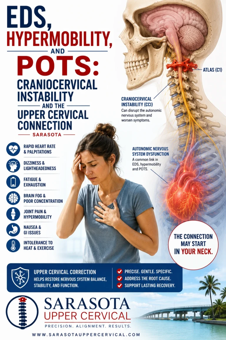
EDS, Hypermobility, and POTS: Craniocervical Instability Sarasota
If you have hypermobile Ehlers-Danlos syndrome or a hypermobility spectrum disorder and you also have POTS, you already know these two things travel together. What you may not have been told is that there is a third structure sitting between them, and it is the one part of the picture most likely to have gone unexamined.
Schedule Your Appointment
Schedule appointmentThe craniocervical junction — the joint complex where the skull meets the atlas and the atlas meets the axis — is stabilized almost entirely by ligament. Unlike the lumbar spine, which has large interlocking facet surfaces and substantial bony containment, the top of the neck achieves its extraordinary range of motion by giving up bony stability and delegating the job to soft tissue. In a connective tissue disorder that alters the behavior of ligament throughout the body, this is the joint with the least margin for error.
This article covers what craniocervical and atlantoaxial instability actually are, what the neurosurgical literature has found about their relationship to autonomic symptoms, the neurological mechanism connecting the upper neck to orthostatic regulation, and — importantly — where conservative care is and is not appropriate. That last section matters more here than in any other article on this site, and if you read only one part of this, read that one.
Definitions worth getting right
Craniocervical instability (CCI) refers to excessive motion between the skull and the upper cervical spine — at the occiput-C1 and C1-C2 levels — resulting from insufficient ligamentous restraint. Atlantoaxial instability (AAI) is the related condition occurring specifically at C1-C2.
The relevant anatomy is small and specific. The transverse ligament runs behind the odontoid process of C2 and holds it against the anterior arch of C1, preventing the atlas from sliding forward on the axis. The alar ligaments run from the odontoid upward to the occipital condyles and limit rotation and lateral bending. The tectorial membrane provides an additional restraint posteriorly. Together these tissues are what stop the head from moving further on the neck than it should. There is no substantial bony lock backing them up.
In hypermobile EDS, connective tissue behaves differently, and these ligaments are subject to the same laxity affecting joints elsewhere. The consequence is not that the head falls off. The consequence is that motion at the craniocervical junction exceeds physiological range in ways that are difficult to detect and that have neurological rather than merely mechanical effects.
Gensemer and colleagues at the Medical University of South Carolina published a 2024 review in Frontiers in Neurology addressing exactly this territory, noting that <cite index="65-1">hEDS patients may experience spinal neurological complications including cervico-medullary symptoms arising from cranio-cervical and cervical instability or hypermobility, as well as tethered cord syndrome — a condition that is often radiographically occult and not always detectable on standard imaging. The point about occult findings applies throughout this discussion and is a recurring theme in why patients go undiagnosed for years.
The symptom picture attributed to cervical instability from ligament laxity is broad and overlapping — headaches, vertigo, tinnitus, vision changes, syncope, radiculopathy, neck pain, and difficulty swallowing appear across the clinical literature. Notice how thoroughly that list overlaps with severe POTS. This is the core diagnostic problem: the two conditions produce substantially the same complaints, and distinguishing them requires deliberately looking for the structural one.
If you developed CCI after whiplash trauma read more here.
Related article
Hypovolemic POTS: Low Blood Volume, Blood Pooling, and the Upper Cervical Connection | Sarasota
Jul 15, 2026What the neurosurgical literature found
The most directly relevant work comes from Fraser Henderson Sr. and colleagues, and it is worth understanding precisely because it is the evidence most often summarized loosely.
Henderson, Rowe, Narayanan and colleagues published a study in World Neurosurgery in 2021 titled "Refractory Syncope and Presyncope associated with atlantoaxial instability: preliminary evidence of improvement following surgical stabilization." The population was patients with instability at the C1-C2 level whose syncope and presyncope had persisted despite maximal medical management. Following surgical stabilization of that junction, autonomic symptoms improved.
That finding is important, and it is important to be precise about what it does and does not show.
What it demonstrates is that the structural integrity of the craniocervical junction can meaningfully affect autonomic function. These were patients whose fainting had not responded to the full range of medical therapy, and the intervention that changed their trajectory was mechanical stabilization of the upper neck. That is a genuinely significant observation about the relationship between this region and autonomic regulation, and it is the strongest single piece of evidence connecting the two.
What it does not demonstrate is that chiropractic care produces comparable outcomes. This was occipito-cervical fusion — a major neurosurgical procedure with substantial risks and permanent loss of motion, performed on a highly selected population with documented instability refractory to everything else. It is not evidence for conservative care. Presenting it as though it were would be dishonest, and it is a leap that appears frequently in marketing material for this condition.
Henderson's group also published longer-term work on cervical medullary syndrome secondary to craniocervical instability and ventral brainstem compression in hereditary hypermobility connective tissue disorders, with five-year follow-up after craniocervical reduction, fusion and stabilization, in Neurosurgical Review. The broader body of work establishes that this region matters. It does not establish what should be done about it in patients who are not surgical candidates.
It is also worth knowing that this whole area is genuinely contested within medicine. Mao and colleagues published a paper in The Spine Journal in 2022 explicitly framed around controversies in the diagnosis and management of craniocervical instability in EDS. Diagnostic thresholds, imaging criteria, and surgical indications are all subjects of active disagreement among specialists. Anyone presenting this field as settled is misrepresenting it.
The neurology connecting the upper neck to orthostatic regulation
Set aside frank structural instability for a moment, because most people with hypermobility and POTS do not have surgical-grade CCI. There is a separate and more broadly applicable mechanism worth understanding.
Blood pressure regulation on standing is a brainstem operation. Baroreceptors in the carotid sinus and aortic arch detect vessel wall stretch and signal through the glossopharyngeal and vagus nerves into the nucleus tractus solitarius in the dorsolateral medulla — the first central relay for cardiovascular afferent information and the integrating hub of the baroreflex. From the NTS, projections run to the caudal ventrolateral medulla, which is inhibitory, and the CVLM restrains the rostral ventrolateral medulla, the principal generator of sympathetic outflow to the heart and vasculature via the intermediolateral cell column. In parallel, the NTS drives cardiac vagal output through the nucleus ambiguus and the dorsal motor nucleus of the vagus.
That circuit is reactive — it responds after pressure has begun to change. Efficient orthostatic regulation also requires anticipation, and anticipation requires knowing that you are standing up. That means knowing where your head is relative to your body.
Two systems supply that information. The vestibular apparatus reports head motion in space. The neck reports head position on the trunk. Both are needed, because the brain must distinguish a whole-body postural change, which requires a cardiovascular response, from a head movement on a stationary body, which does not. Tilting your head back to look at a ceiling produces vestibular signals resembling those of standing, but your legs require no vasoconstriction because your body has not moved.
Bolton, Kerman, Woodring and Yates demonstrated in 1998 that stimulating the C2 dorsal root ganglion and the C2 and C3 branches supplying the dorsal neck muscles produced measurable responses in splanchnic sympathetic nerve activity, and showed that cervical and vestibular influences on sympathetic output behave antagonistically — precisely the arrangement required for that discrimination. Human research has since shown that stretching neck muscles modulates muscle sympathetic nerve activity directed to the lower limbs, with the investigators concluding that neck proprioceptors may contribute to blood pressure regulation during orthostatic challenge.
The sensor supplying this information is the suboccipital muscle group — rectus capitis posterior major and minor, obliquus capitis superior and inferior — which carries one of the highest muscle spindle densities found anywhere in the human body. These are short muscles crossing one or two joints whose primary role is not force production but continuous, high-resolution reporting of head position. Rectus capitis posterior minor additionally connects to the spinal dura through the myodural bridge. The vagus nerve itself exits the skull through the jugular foramen and descends within the carotid sheath immediately anterior to the transverse process of the atlas.
Now apply that to a hypermobile joint. Proprioceptive receptors report joint position by detecting tissue tension. In a joint whose ligamentous restraints are lax, the relationship between actual position and tissue tension is altered — the signal reaching the brainstem may be less accurate, less consistent, or asymmetric between sides. The system responsible for inferring body position from head position is working from degraded input, and the cardiovascular response that depends on that inference is correspondingly less well calibrated.
This is a hypothesis. It is built on documented physiology — the cervicosympathetic pathway is real, the spindle density is real, the anatomy is real — but the specific claim that upper cervical proprioceptive degradation contributes to POTS in hypermobile patients has not been demonstrated in controlled research, and it should be held as a plausible mechanism rather than an established fact.
It is also worth saying clearly that the neck is not the only route from EDS to POTS, and probably not the main one. Connective tissue laxity affects blood vessels directly. Lax vasculature distends more readily under gravitational load, permitting greater venous pooling in the legs and abdomen on standing. That mechanism requires no involvement of the cervical spine whatsoever and is likely a major driver of orthostatic intolerance in this population. Reduced blood volume and the frequently co-occurring mast cell activation add further layers. Anyone framing the neck as the explanation for POTS in EDS is overreaching.
Where conservative care fits — and where it does not
This section is the reason this article needed to be written carefully.
True structural craniocervical instability is a medical diagnosis, and it changes what is safe. High-velocity cervical manipulation is not appropriate in the presence of frank instability at the craniocervical junction. That is not a cautious hedge; it is a genuine contraindication, and it is the reason that anyone with hypermobility, suspected CCI or AAI, or a connective tissue diagnosis needs proper evaluation before any hands-on cervical care — including ours.
Related article
Thoracic Outlet Syndrome Treatment and Relief Sarasota | Sarasota Upper Cervical
Apr 28, 2026Imaging deserves specific mention because the standard workup routinely misses this. A supine MRI and a neutral X-ray are taken in exactly the position in which an unstable joint appears normal, because the instability only manifests under load or at end range. Assessment for atlantoaxial instability involves examining rotation of C1 on C2, vertical displacement measured against the Chamberlain, McRae and McGregor lines, and horizontal displacement via the atlanto-dens interval; craniocervical instability assessment uses measures including the Grabb-Mapstone-Oakes measurement. Upright and dynamic imaging is often required. It is entirely possible to be told your imaging is normal and still have significant instability, because the imaging performed was not designed to detect it.
Where conservative management does have a role, the literature points toward stabilization rather than mobilization. Russek and colleagues published work in Frontiers in Medicine in 2023 on the presentation and physical therapy management of upper cervical instability in patients with symptomatic generalized joint hypermobility, and the general principle in that work is retraining the deep cervical stabilizers and improving proprioceptive control — building active support for a joint whose passive support is compromised. The instinct to loosen a stiff-feeling neck is often exactly wrong in this population, because the stiffness is frequently protective guarding around a joint the body does not trust.
For upper cervical chiropractic specifically, the appropriate approach in hypermobile patients is low-force and precision-based rather than manipulative, and it should only follow an evaluation that has actively assessed for instability rather than assumed its absence. Some hypermobile patients are reasonable candidates for this care. Some are not, and should be referred for neurosurgical or neurological evaluation instead. Distinguishing between those two groups is the single most important thing a clinician does in this population, and any practice that does not take that distinction seriously should be avoided.
The honest position on evidence is that upper cervical chiropractic research in POTS consists of case reports and small series, including a 2024 Cureus case report by Trager and colleagues describing marked improvement in a patient with cervicogenic dizziness, upper cervical instability and POTS. Case reports cannot establish causation and their outcomes should not be presented as typical. There are no controlled trials of upper cervical chiropractic care in hypermobile patients with POTS.
What to do with this
If you have hypermobility and POTS, the medical foundation comes first, and it is not optional. That means physician-directed autonomic management, and where symptoms suggest cervico-medullary involvement — progressive neurological signs, myelopathic symptoms, severe positional headache, difficulty swallowing, or syncope refractory to management — it means neurological or neurosurgical evaluation with appropriate upright dynamic imaging. Those symptoms warrant medical assessment, not conservative care.
If your picture is orthostatic intolerance with upper neck pain, suboccipital tightness, headaches, and position-dependent dizziness, without progressive neurological signs, then an upper cervical evaluation is a reasonable component of a broader workup — with the explicit understanding that its first job is to determine whether care is appropriate for you at all.
The thing worth holding onto is that in this population the craniocervical junction is not a peripheral consideration. It is the joint in the body with the least bony stability and the most neurological consequence, in people whose ligaments behave differently, who overwhelmingly also have autonomic dysfunction. It deserves to be looked at properly — carefully, with the right imaging, by someone who knows when to refer.
If you have hypermobility or EDS along with POTS and want a careful evaluation of whether your upper cervical spine is contributing — including an honest assessment of whether conservative care is appropriate in your case — schedule a free consultation or call 941-259-1891.
What do upper cervical chiropractors do?
Upper cervical chiropractic is a distinct discipline within chiropractic, and it does not look like what most people picture. There is no twisting of the head, no pulling on the neck, and no forceful popping or cracking. Instead, care begins with measurement. Three-dimensional cone beam CBCT imaging is used to map the precise alignment relationship between the skull, the atlas, and the axis, so that any correction is calculated for your individual anatomy rather than delivered by feel. Functional leg length assessment is used alongside imaging as a postural indicator, checked before and after care to monitor how the body is responding. The correction itself is low-force and specific, directed along a calculated vector rather than applied broadly to the spine. And the underlying principle is the opposite of what people expect from a chiropractor: the aim is not to adjust you repeatedly, but to make a precise correction and then let it hold. When alignment holds, the nervous system is given a stable environment to work in, and adjustments become less frequent over time rather than more. Holding the correction is the goal — not the number of visits.
Medical disclaimer: This article is for educational purposes only and does not constitute medical advice, diagnosis, or treatment. Craniocervical and atlantoaxial instability are medical diagnoses requiring evaluation by qualified physicians, and cervical manipulation may be contraindicated in their presence. Anyone with a connective tissue disorder, hypermobility, or suspected cervical instability should obtain appropriate medical evaluation before undertaking any hands-on cervical treatment. Upper cervical chiropractic care is a complementary approach and is not a substitute for medical or surgical management. Individual results vary, and outcomes described in published case reports and surgical studies should not be interpreted as typical or applicable to conservative care. Always consult your physician before making changes to your care.


Leave a comment