

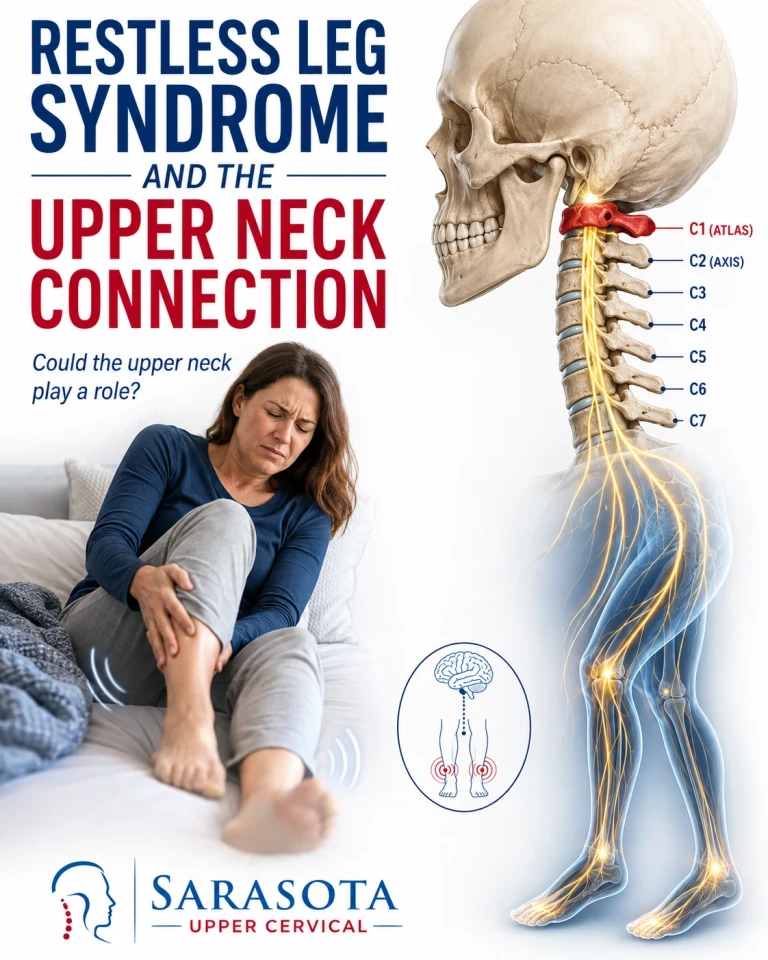
Restless leg syndrome treatment in Sarasota, Bradenton, and Lakewood Ranch usually focuses on the legs, which is understandable, since that is where the sensation lives. But the legs are not generating it. Restless leg syndrome is a disorder of how the nervous system inhibits movement, and the machinery that does the inhibiting sits in the brain and travels down the spinal cord. The upper neck is where all of it passes through.
What the diagnosis actually requires
Schedule Your Appointment
Schedule appointment
Restless leg syndrome is diagnosed clinically on four features: an urge to move the legs, usually with uncomfortable sensations; symptoms that begin or worsen at rest; relief with movement; and a clear evening or night-time worsening. The circadian pattern is not incidental. It is diagnostic, and it points toward a regulatory system rather than a structural leg problem.
Rule out iron first
Before any structural discussion, one thing has to happen. Brain iron deficiency is the most robustly established finding in restless leg syndrome, and serum ferritin can be low while a standard blood count looks normal. Current guidance recommends checking ferritin and considering iron repletion when it falls below roughly 75 nanograms per millilitre. Kidney disease, pregnancy, peripheral neuropathy, and several common medications — antihistamines, many antidepressants, and antinausea drugs that block dopamine — also cause or aggravate it.
If your ferritin has never been checked, that is the first appointment to make, not this one.
The spinal cord is hyperexcitable — this part is measured, not theorised
Here the evidence is genuinely strong, and it is the core of the argument.

People with restless leg syndrome demonstrate measurable spinal hyperexcitability on electrophysiological testing. The findings include exaggerated crossed-extensor reflexes, a hyperexcitable plantar reflex, an altered cutaneous silent period, and deficits in presynaptic inhibition, post-activation depression, reciprocal inhibition, and recurrent inhibition. Gemignani and colleagues, writing in the Journal of Sleep Research in 2018, framed the condition as a flexor reflex circuitopathy on exactly this basis.
Read that list again. Every item is a failure of inhibition. The spinal cord is not producing new signals; it has lost the braking that normally suppresses them. Reviewers attribute this to several factors: reduced descending dopaminergic input, dysfunction of inhibitory interneurons including Renshaw cells, sensitisation of spinal central pattern generators, and sensitisation of the dorsal root ganglia.
Where the brake comes from — and why it involves the autonomics
The principal source of dopamine reaching the spinal cord is the A11 cell group, which sits in the posterior hypothalamus and sends the diencephalospinal pathway the entire length of the cord. It is worth being precise: A11 is diencephalic, not brainstem. But its fibres, along with the corticospinal and cerebello-rubro-spinal tracts, descend through the brainstem and the upper cervical cord to reach the lumbar segments where restless leg symptoms are expressed.
A11 projects most strongly to two places: the sensory dorsal horn, and the intermediolateral nucleus — the column of sympathetic preganglionic neurons. Clemens and colleagues, in Neurology in 2006, argued that A11's action on those sympathetic neurons is inhibitory, so losing it shifts the balance toward sympathetic excitation. Increased sympathetic drive raises circulating catecholamines, which aberrantly activate high-threshold muscle afferents in the leg, which is felt as the urge to move.
That is a coherent account of why restless leg syndrome is simultaneously a sensory disorder, a motor disorder, and an autonomic one — and why periodic limb movements come with surges in heart rate and blood pressure.
The finding that argues against the simple version
I am not going to leave this out. Earley, Allen, Connor, Ferrucci, and Troncoso examined A11 dopaminergic neurons in autopsy brains from people with restless leg syndrome and found them structurally normal. A11 damage has never been demonstrated in humans. The dopamine hypothesis rests largely on the indirect evidence that dopaminergic drugs relieve symptoms.
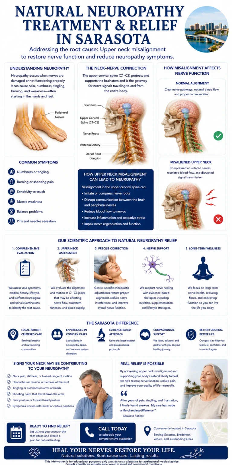
That is an important qualification, and it points somewhere useful. If the cells are intact, the problem may lie in the function of the pathway rather than the survival of the neurons — and function depends on the route the fibres travel.
The upper cervical spine as the common corridor
Every descending inhibitory tract implicated in restless leg syndrome converges through the same anatomical bottleneck: the transition from brainstem to spinal cord at the level of the atlas.
Two lines of evidence make this more than geography. First, restless leg syndrome and periodic limb movements are documented in association with myelopathy, reported by Yokota and colleagues in 1991 and Hartmann and colleagues in 1999. Second, and more strikingly, Salminen and colleagues showed in 2013 that periodic limb movements can persist below a complete lesion of the cervical spinal cord — consistent with a spinal generator being released when descending inhibition is interrupted. In animal work, discrete lesions of the corticospinal tract made at the C1 level produced restless-leg-like movements during sleep.
So interruption of descending inhibition at the upper cervical level can produce these symptoms. That is established.
What is not established is that atlas misalignment does the same thing. Those studies involved complete cord lesions and surgical ablation, not subtle positional change, and there is no clinical trial of upper cervical chiropractic for restless leg syndrome. The reasoned proposition — that upper cervical dysfunction may alter the mechanical and afferent environment of a corridor already implicated in this condition — is a hypothesis built on real anatomy. I would rather label it that way than pretend it is proven.
Precision, and knowing when not to adjust
Because the reasoning is anatomical, the correction must be too. Cone beam computed tomography measures the three-dimensional relationship between skull, atlas, and axis against your individual anatomy, so any correction is calculated rather than estimated. There is no twisting, no popping, no cracking, no pulling of the neck.
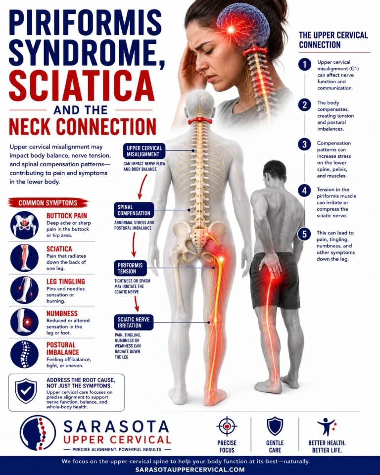
The goal is not frequent adjusting. It is holding. A correction that stays in place allows the nervous system a sustained period of unchanged input, which is where any adaptive change would occur. Repeatedly adjusting a spine that is already holding adds mechanical insult without benefit.
This is why objective testing governs the decision. Paraspinal infrared thermography tracks skin temperature differentials as a proxy for autonomic regulation, and the question asked is whether a patient's pattern has stabilised, not whether it matches a normal chart. Leg length assessment, balance and postural measurement, and cervical range of motion are taken before and after and at every subsequent visit.
When those measures indicate the correction is holding, no adjustment is given. That is the more important half of the protocol, and it is the part most patients are never told about.
Red flags
Progressive leg weakness, numbness in a saddle distribution, changes in bladder or bowel control, unsteady gait, or symptoms in the arms as well as the legs suggest spinal cord involvement and require prompt medical evaluation rather than manual care. New restless leg symptoms alongside unexplained weight loss, fever, or a history of cancer need medical work-up. Symptoms that are constant rather than worse in the evening, or that do not improve with movement, are probably not restless leg syndrome and deserve a different diagnosis.
An evaluation in Sarasota
If your ferritin has been checked, secondary causes have been addressed, and symptoms persist — particularly if they began after a head or neck injury, or if you have upper neck pain or dizziness alongside them — an upper cervical evaluation can determine whether the craniocervical junction is a contributing factor. This works alongside your physician and any sleep or neurological care rather than in place of it. To arrange a consultation, visit NeckWise.
Dr. Drew Hall, Upper Cervical Chiropractor
Serving Sarasota, Bradenton, and Lakewood Ranch, Florida
This article is for general educational purposes only and is not medical advice, a diagnosis, or a treatment recommendation for any individual. Restless leg syndrome should be diagnosed and managed by a qualified medical provider, and evaluation should include serum ferritin and a review of contributing medications and secondary causes. Upper cervical chiropractic is an area of focus within chiropractic; it is not a board-recognized specialty, and no claim of specialization or superiority is made or implied. No clinical trial evidence exists for upper cervical chiropractic care in restless leg syndrome, and none is claimed here; the anatomical relationship described is presented as a reasoned hypothesis rather than an established treatment mechanism. Cone beam computed tomography is used as a precision measurement and analysis tool, not as a stand-alone diagnostic device, and thermography and postural measures are used to track change within an individual over time rather than to diagnose disease. Medication decisions belong solely with your prescribing physician. Individual results vary and no specific outcome is guaranteed. Seek prompt medical attention for progressive weakness, numbness, changes in bladder or bowel control, or unsteady gait. Always consult a qualified healthcare provider regarding diagnosis and treatment.


Leave a comment