
How 3D imaging of the joints between your head and neck guides a correction built for your anatomy alone
In upper cervical care, precision is everything. The difference between a correction that holds and one that does not often comes down to a single question: do we actually know how this particular person's spine is built? At Sarasota Upper Cervical, the tool that answers that question is CBCT — cone beam computed tomography — and the method that turns those images into a precise, individualized correction is the Blair analysis.
Schedule Your Appointment
Schedule appointmentThis guide explains what CBCT is, why being able to see the joints between the head and neck in three dimensions matters so much, how the Blair analysis uses that information, and why knowing your unique anatomy is what makes a gentle, no-twist correction both possible and lasting.
Curious whether a precise upper cervical correction could help you?
Schedule a free consultation to find out whether you are a candidate. Call 941-259-1891 to get started. Or CLICK HERE.
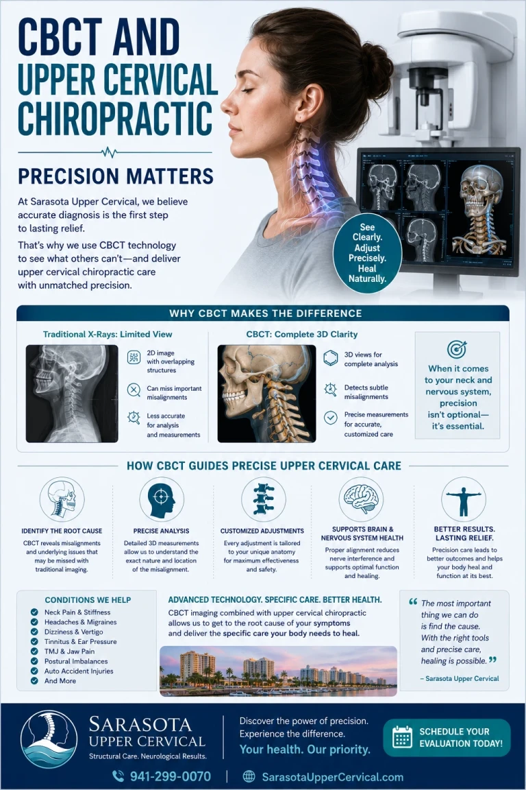
What Is CBCT?
CBCT stands for cone beam computed tomography. It is an advanced imaging technology that captures the structures of your upper neck as a true three-dimensional volume rather than a flat picture. A traditional X-ray collapses everything in its path onto a single two-dimensional plane, layering bones on top of one another so that fine detail is lost or hidden. CBCT instead reconstructs the region in 3D, so the individual joints, surfaces, and angles can be viewed from any direction.
For the upper cervical spine, this is not a luxury — it is essential. The joints between the skull and the atlas (C1), and between the atlas and the axis (C2), are small, deep, intricately shaped, and oriented at angles that differ from person to person. A flat image simply cannot show their true orientation. CBCT can. It is used here strictly as a precision measurement tool — a way to see and measure your anatomy accurately — so that any correction is based on your real structure rather than an assumption.
Why Seeing the Joints Between the Head and Neck Matters
Most people assume one neck is built much like another. In reality, the joints at the very top of the neck are remarkably individual. The joint between the skull and the atlas is not a flat, planar joint like most of the spine. It is cup-shaped — the skull rests in the atlas almost like an object cradled in a holder. This cradle shape, and the exact angles of the joint surfaces, vary from one person to the next.
That individuality is the entire reason imaging matters. If you cannot see the actual shape and angle of a person's joints, you cannot know how their spine has misaligned, and you are left to guess. Seeing the joints directly removes the guesswork. It lets the doctor understand the specific architecture of your upper neck before deciding anything about correction.
There is also a broader benefit to seeing clearly: the true source of a problem is often not where the symptoms are. By viewing the whole region in detail, care can focus on the actual primary misalignment rather than chasing the spot that happens to hurt.
The Three-Dimensionality of Misalignment
Here is one of the most important and least understood ideas in upper cervical care: when the atlas misaligns, it does not simply slide straight to one side. Because the joint surfaces are cupped and angled, the atlas moves along a three-dimensional path determined by the shape of those joints.
Related article

Before You Get Ear Tubes or Balloon Dilation: Conservative Options for Eustachian Tube Dysfunction
Jun 30, 2026Dr. William Blair, who developed the Blair technique, described this decades ago using the idea of a track. Picture the two joint surfaces as a pair of railroad tracks. In a healthy neck they run in parallel and the head moves smoothly along them in a normal nodding motion. After an injury, it is as if the tracks no longer line up — and as the "train" moves, one side stays on its track while the other slips off. The atlas tracks along one joint while sliding off the other, producing a misalignment that is genuinely three-dimensional, following the unique geometry of that person's joints.
This model was once theoretical. Today, modern CBCT imaging confirms it: the atlas does misalign along a 3D path dictated by the shape of the joints, exactly as Blair proposed. This matters enormously for correction, because if the misalignment happened in three dimensions along a specific path, then the correction has to be delivered back along that same path — not in some generic sideways direction. You can only know that path if you can see the joints in three dimensions.
The Blair Analysis: Turning Images Into an Individualized Plan
The Blair analysis is the method used to study your CBCT images and translate them into a precise correction plan built for you. Its founding insight is profound in its simplicity: because everyone's spine is naturally asymmetric, you cannot rely on generic landmarks or assume that the left and right sides are mirror images. You have to measure each person's actual anatomy.
This is exactly why Blair developed his approach. Trained originally in earlier upper cervical methods, he became concerned that natural asymmetry — the normal variation in the shape of the bones from one person and one side to the next — could make standard analysis inaccurate. If a vertebra is simply shaped differently on one side, that difference could be mistaken for a misalignment that is not really there, or could hide one that is. The Blair analysis accounts for this by studying your individual joint orientations directly.
Using carefully positioned CBCT images, the Blair analysis identifies which specific joint has misaligned and the precise angle of that misalignment. As one way of describing it: each person's anatomy is different, so the imaging is used to uncover the blueprint for correcting that individual's unique misalignment pattern. No two blueprints are identical, because no two spines are.
Why Individual Anatomy Determines the Path of Misalignment
It is worth stating this central principle plainly, because it ties everything together: the way your spine misaligns is dictated by the way your spine is built.
The angles of your joint surfaces, the shape of your condyles where the skull meets the atlas, the formation of your atlas and its transverse processes — all of these are unique to you, and all of them determine the direction and pattern in which your atlas can move when it misaligns. Two people who experience a similar injury can end up with very different misalignment patterns simply because their joints are shaped differently. This is why a one-size-fits-all approach falls short, and why knowing your specific anatomy is paramount. Without that knowledge, even a well-intentioned correction is an educated guess. With it, the correction can be matched precisely to how your spine actually moved out of alignment.
Finding the Proper Contact Point on the Atlas
Precision is not only about knowing the angle of correction — it is also about knowing exactly where to make contact. The correction is delivered at a specific point on the atlas, most often its transverse process (the small bony projection on the side of the vertebra). But here again, anatomy is individual: the transverse processes themselves vary in their formation and are not always perfectly symmetric from side to side.
This is where seeing the anatomy on imaging becomes invaluable. Rather than relying solely on feeling for a landmark through the skin — which research has shown can be misleading precisely because of natural asymmetry — the doctor uses the imaging to understand the true position and shape of your transverse process. Using anatomical landmarks identified on your own CBCT, the contact can be placed exactly on the correct part of the transverse process of the atlas, on the correct side, at the correct angle. Getting precisely onto the right structure is what allows a light, specific correction to do its work.
Precision Enables a Gentle Correction — No Popping, Twisting, or Yanking
People are often surprised to learn that a more precise technique is also a gentler one. The two go hand in hand. When you know exactly which joint has misaligned, in which direction, and exactly where to contact it, you no longer need force. You do not need to twist the neck, crank it, or produce a popping sound. You simply need to apply a light, specific correction along the precise path the analysis revealed.
Blair upper cervical corrections involve no twisting, popping, or yanking of the neck. The adjustment is gentle and exact. This is possible only because of the precision that comes first — the imaging and analysis do the demanding work of figuring out the what, where, and how, so that the correction itself can be delivered softly. For patients whose nervous systems are already sensitized, this gentle approach is not just more comfortable; it is more appropriate.
The Principle of Holding the Adjustment
There is a principle at the heart of Blair upper cervical care that reframes what the correction is even for: it is not the act of adjusting that produces healing — it is the body holding its proper alignment over time that allows healing to occur.
In other words, the goal is not to be adjusted again and again. The goal is to make a precise correction and then have your body hold that correction — to stay in alignment. When the spine holds, the nervous system is given a stable, uninterrupted environment in which to recover and regulate. That is when meaningful change tends to happen.
This is also why each visit involves objective testing rather than an automatic adjustment. The doctor checks whether you are still holding your correction. If you are, no adjustment is needed that day — and that is a good thing, a sign of stability. If testing indicates the correction has shifted, a precise adjustment is made again. Over time, as the spine learns to hold, corrections are typically needed less and less often. Precision is what makes holding possible in the first place: a correction matched accurately to your anatomy is far more likely to stay than a generic one.
How Precision Leads to Better Outcomes
Bring these threads together and the logic is clear. Seeing the joints between your head and neck in three dimensions reveals the real, individual shape of your spine. The Blair analysis uses that 3D detail to determine exactly which joint misaligned and along what path, accounting for your natural asymmetry. Knowing the path and the proper contact point allows a gentle, specific correction with no twisting or popping. And a correction matched precisely to your anatomy is the kind most likely to hold — which is what gives your nervous system the stable conditions it needs.
Precision is not a technical detail; it is the entire foundation of better, more lasting results. The more accurately the correction fits your unique structure, the better the chance your body holds it and responds. That is the case for doing this carefully, with imaging and individualized analysis, rather than by assumption.
A Note on Candidacy
Not everyone's health concerns trace back to the upper cervical spine, and not everyone is a candidate for this care. That is part of why a thorough evaluation — including appropriate imaging — comes first. The assessment is designed to determine whether your specific anatomy and alignment show the structural signs that this approach addresses, and to tell you honestly if they do not. Precision applies to candidacy too: the goal is to help the people this care can genuinely help.
Curious whether a precise upper cervical correction could help you? Schedule a free consultation to find out whether you are a candidate. Call 941-259-1891 to get started. Or book here.
Frequently Asked Questions
What is CBCT, and how is it different from a regular X-ray?
CBCT (cone beam computed tomography) captures your upper neck as a true three-dimensional volume, rather than the flat, single-plane image a standard X-ray produces. This lets the doctor see the actual shape and orientation of the small joints between your skull, atlas, and axis from any angle — detail that a flat image cannot reveal. It is used as a precision measurement tool to guide a correction based on your real anatomy.
Why does seeing the joints in 3D matter for my care?

The joints at the top of the neck are cup-shaped and individually angled, so when the atlas misaligns it moves along a three-dimensional path determined by the shape of your joints. Seeing that path in 3D is what allows the correction to be delivered precisely back along it, rather than in a generic direction. You can only know the path if you can see the joints.
What is the Blair analysis?
The Blair analysis is the method used to study your CBCT images and build an individualized correction plan. Its core principle is that everyone's spine is naturally asymmetric, so generic assumptions can be misleading. By studying your specific joint orientations, the analysis identifies which joint has misaligned and at what precise angle — a blueprint unique to you.
How can the correction be effective without twisting or popping?
Precision is what removes the need for force. Once imaging and analysis reveal exactly which joint misaligned, in which direction, and exactly where to make contact on the atlas, the correction can be delivered as a light, specific input along that path — no twisting, popping, or yanking. The demanding work happens in the analysis, so the adjustment itself can be gentle.
What does "holding the adjustment" mean?
In Blair upper cervical care, it is not the act of adjusting that produces healing — it is your body holding its proper alignment over time. The goal is to make a precise correction and have your spine stay in alignment, giving the nervous system a stable environment to recover. Each visit includes objective testing; if you are holding, no adjustment is made that day. As the spine learns to hold, corrections are usually needed less often.
How do you know exactly where to make the correction?
The contact is typically made on the transverse process of the atlas, but these vary in shape and symmetry from person to person. Using anatomical landmarks identified on your own CBCT images, the doctor can place the contact precisely on the correct part of the transverse process, on the correct side and angle, rather than relying only on feeling for a landmark through the skin, which natural asymmetry can make misleading.
How do I find out if this is right for me?
The first step is a consultation and evaluation, which may include CBCT imaging to assess your individual anatomy and alignment. This determines whether your case shows the structural signs that upper cervical care addresses.
If it does not, you will be told so. Call 941-259-1891 to schedule a free consultation. Or book here.
This content is for informational purposes only and does not constitute medical advice. CBCT is used as a precision imaging tool to guide individualized upper cervical care; candidacy is determined by evaluation, and individual results vary.


Leave a comment