

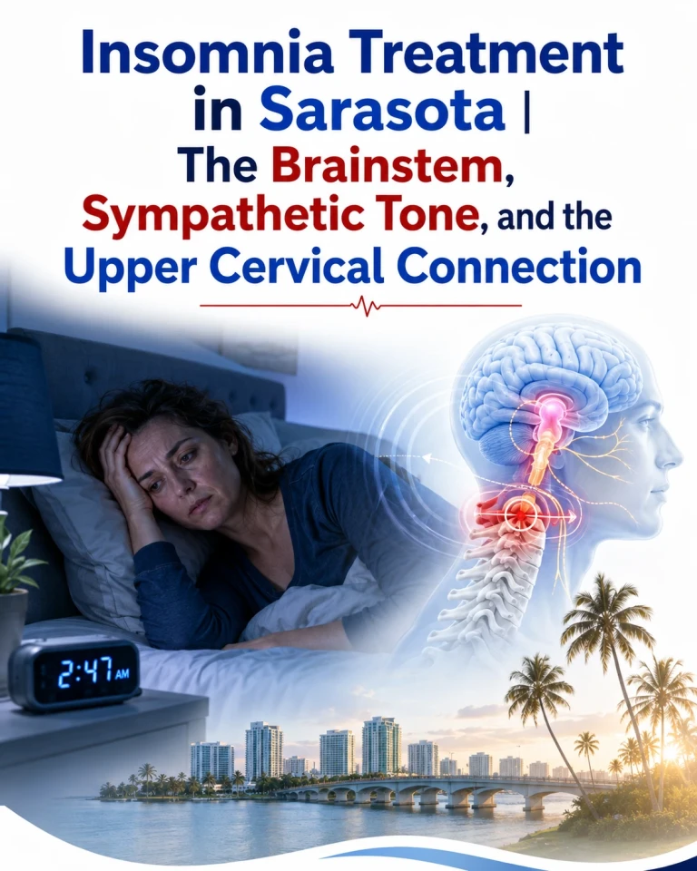
Insomnia treatment in Sarasota usually begins and ends with the same two options: a sleep hygiene handout and a prescription. For a large number of people those options help, and if they have worked for you, there is no reason to look further. But there is a specific group of patients for whom neither one ever seems to move the needle, and they tend to share a history that nobody in the sleep conversation ever asked about — a car accident, a fall, a concussion, a wrestling match, a hard tackle. This article is about the neurological pathway that may connect that old injury to the fact that you cannot shut your brain off at eleven at night, or you wake up in the morning feeling nonrestored sleep. and We will also discuss what honest, objectively measured upper cervical care can and cannot claim in that conversation.
The Problem Usually Isn't Sleep Drive — It's the Inability to Downshift
Schedule Your Appointment
Schedule appointment
One of the more useful reframes in modern sleep medicine is that chronic insomnia is not primarily a deficiency of sleepiness. It is an excess of arousal. The dominant explanatory framework in the research literature, generally credited to Dieter Riemann and colleagues and refined over the last fifteen years, is called the hyperarousal model. It holds that people with insomnia disorder are not tired-but-unable-to-sleep so much as they are physiologically switched on around the clock, day and night, in a way that good sleepers are not.
The evidence for this is not soft. Bonnet and Arand, working in the late 1990s, found elevated heart rate in patients with insomnia compared with matched controls, along with a heart rate variability signature — increased low-frequency power and decreased high-frequency power — that is generally interpreted as a shift toward sympathetic dominance. Vgontzas and colleagues, sampling plasma cortisol every thirty minutes across a full twenty-four hours, found significantly elevated cortisol in insomnia patients relative to good sleepers. Later work has added increased core body temperature, elevated whole-body metabolic rate, higher global cerebral glucose metabolism, and an increase in fast-frequency EEG activity in the beta and gamma bands that persists into sleep itself. This is established physiology, replicated across multiple independent laboratories, and it is not seriously contested.
What it means practically is this. If your nervous system is running with the sympathetic branch idling high, the problem is not that you need more sleep pressure. You already have plenty. The problem is that the brake is not engaging. And that reframe matters enormously, because sleep hygiene advice and sedative medication both address the wrong end of the equation — one tries to increase sleep drive, the other tries to chemically override arousal. Neither asks why the arousal is elevated in the first place.
How the Brainstem Actually Switches Sleep On and Off
Sleep is not something the brain drifts into. It is actively imposed by a specific piece of circuitry. The ventrolateral preoptic nucleus in the anterior hypothalamus contains GABAergic and galaninergic neurons whose job is to inhibit the wake-promoting centers. Those wake-promoting centers — the ascending reticular activating system, the orexin/hypocretin neurons of the lateral hypothalamus, the noradrenergic locus coeruleus in the pons, the serotonergic raphe nuclei, the histaminergic tuberomammillary nucleus — inhibit the ventrolateral preoptic nucleus right back. The two sides suppress each other, which produces a bistable switch: the system tends to sit firmly in wake or firmly in sleep rather than lingering in between.
That architecture explains a lot about the phenomenology of insomnia. When the arousal side of the switch carries excess tone, the flip does not complete cleanly. You get the drift toward sleep followed by a lurch back to full alertness. You get micro-arousals, which are among the most consistently replicated findings in insomnia polysomnography. You get the subjective experience patients describe over and over in our Sarasota office — being exhausted and wired at the same time.
The locus coeruleus deserves particular attention here, because it is the brain's principal source of noradrenaline and it sits in the dorsal pons, immediately adjacent to the upper cervical junction. Locus coeruleus firing drops sharply as sleep deepens and goes essentially silent during REM. It is, functionally, the central node where general arousal and sympathetic tone converge. This is established neuroanatomy taught in every graduate neuroscience curriculum.
The Sympathetic Pathway That Runs Through the Neck
Here is where the anatomy gets genuinely interesting, and where I want to be careful to say precisely what is known and what is not.
Melatonin is not manufactured on a whim. Its release from the pineal gland is controlled by a defined multi-synaptic sympathetic circuit. Light information reaches the suprachiasmatic nucleus of the hypothalamus through the retinohypothalamic tract. The suprachiasmatic nucleus projects to the paraventricular nucleus. The paraventricular nucleus projects down to the intermediolateral cell column of the spinal cord. Preganglionic sympathetic fibers then run to the superior cervical ganglion, and postganglionic fibers from that ganglion ascend to the pineal gland, where noradrenaline acting on beta-1 and alpha-1 adrenoceptors drives melatonin synthesis. This is established physiology, mapped in detail by Robert Moore and others, and confirmed by the observation that surgically removing the superior cervical ganglion abolishes the normal melatonin rhythm.
Two honest caveats belong here, and I would rather state them plainly than let the article imply something it cannot support.
First, the preganglionic cell bodies for this circuit are located in the upper thoracic intermediolateral cell column, not at C1 or C2. So it is simply not true — and you will see it claimed on plenty of chiropractic websites — that an atlas misalignment directly compresses the melatonin pathway. It does not. Second, the superior cervical ganglion does lie anterior to the transverse processes of roughly C1 through C3, which makes it an upper cervical structure by location, but proximity is not the same thing as demonstrated mechanical interference, and I am not aware of imaging or physiological evidence in humans showing that atlas position mechanically compromises that ganglion.
Related article
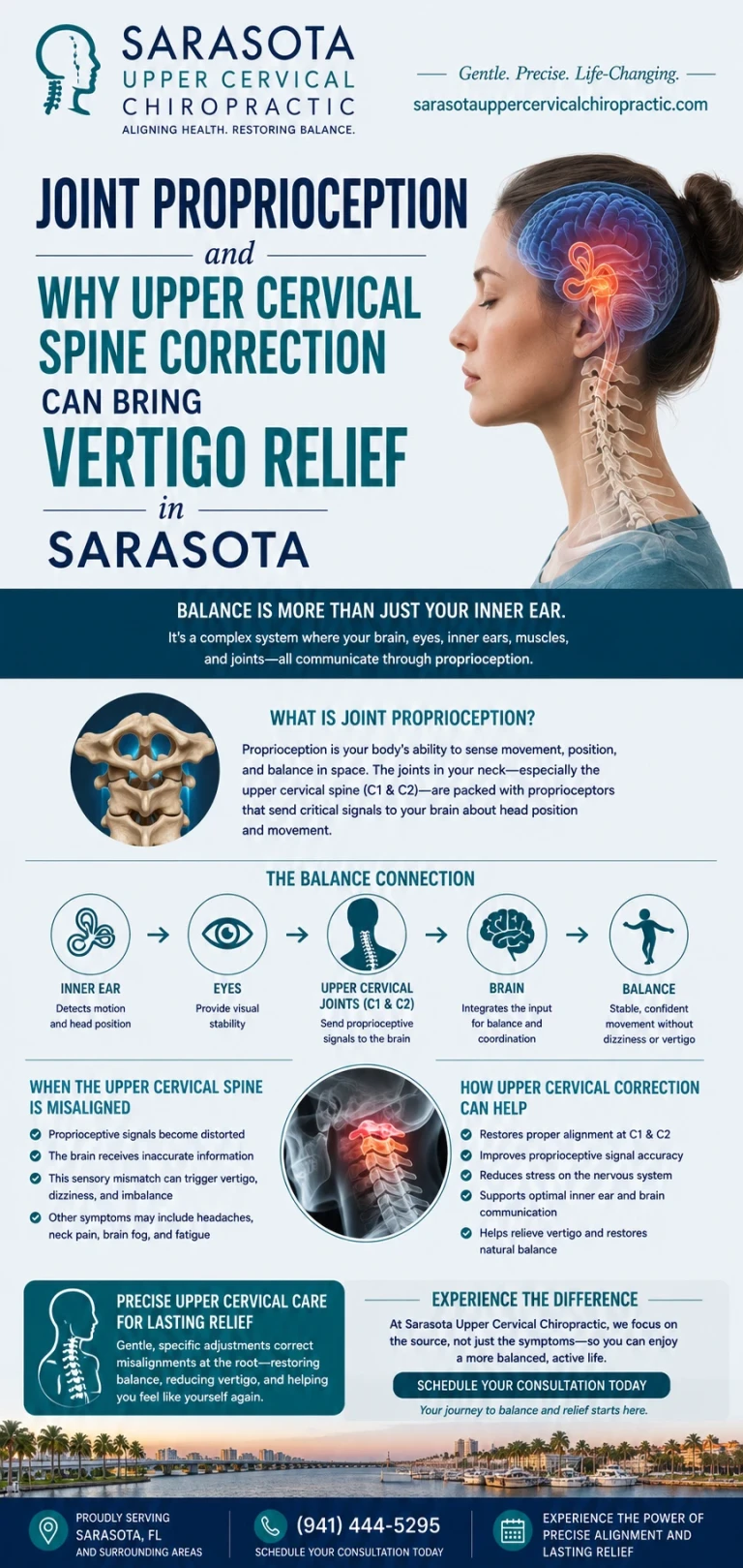
Joint proprioception and why upper cervical spine correction can bring relief in sarasota
Jun 02, 2026What the literature does support is a different and, in my view, more interesting route: upper cervical sensory input modulates sympathetic outflow centrally, through the brainstem, rather than by squeezing a peripheral nerve.
Why the Upper Neck Has an Unusual Amount of Say Over Autonomic Tone
The suboccipital muscles are built like sensory organs rather than movers. Kulkarni, Chandy and Babu, publishing in Neurology India in 2001, quantified muscle spindle density in these muscles and reported 242 spindles per gram in obliquus capitis inferior, 190 in obliquus capitis superior, and 98 in rectus capitis posterior. For comparison, opponens pollicis — a muscle we think of as exquisitely controlled — comes in around 17. The important caveat is that the Kulkarni measurements were taken from stillborn human fetuses, so the absolute numbers should not be transplanted uncritically onto adults, but the qualitative finding that this region carries extraordinary proprioceptive density is corroborated across multiple independent anatomical studies.
The question is whether that dense afferent stream actually reaches autonomic circuitry, and the answer appears to be yes. Bolton, Kerman, Woodring and Yates, publishing in Brain Research Bulletin in 1998, stimulated the C2 dorsal root ganglion and the C2 and C3 nerve branches supplying dorsal neck muscles in anaesthetised cats, and recorded responses in the splanchnic sympathetic nerve as well as in respiratory nerves. Upper cervical afferent input demonstrably changed sympathetic outflow. Later anatomical tracing work by Edwards, Deuchars and colleagues followed those same C2 afferents centrally and found they project to the intermedius nucleus of the medulla, a structure embedded in cardiorespiratory and autonomic control circuitry — and those authors explicitly framed their investigation around the autonomic abnormalities seen in whiplash-associated disorders.
There is also human evidence. Kuwagata and colleagues found that neck flexion increased muscle sympathetic nerve activity, heart rate and blood pressure, and critically, the response persisted in brain-dead patients in whom vestibular contribution was absent — which argues that the neck itself, not just the inner ear, is driving part of that sympathetic response.
Two things need saying about how far this carries. Bolton's work showed that cervical afferent stimulation could either excite or inhibit splanchnic sympathetic activity depending on conditions — it is not a simple dial where more neck input equals more sympathetic tone. And all of this is animal and acute-human physiology. The proposition that a chronic atlas misalignment produces a sustained shift in resting sympathetic tone sufficient to disrupt the sleep switch is a reasoned hypothesis extrapolated from that established physiology — it has not been demonstrated in clinical trials. I think it is a good hypothesis. It is not a proven mechanism, and anyone who tells you otherwise is selling something.
Whiplash, Sports Injury, and the Trauma That Started It
Most patients who arrive at our Sarasota office with treatment-resistant insomnia and a suspicious cluster of accompanying symptoms have a trauma history. Often they have stopped connecting it to anything, because the accident was in 2011 and the sleep fell apart in 2016 and those seem like separate stories.
The injury mechanics are well characterised. Manohar Panjabi and Beth Winkelstein and their respective collaborators demonstrated that in whiplash loading, cervical facet capsular ligaments undergo strains that exceed physiological range but fall short of gross rupture — subfailure injury. That is precisely the category of damage that standard imaging misses: no fracture, no dislocation, normal radiographs, patient told nothing is wrong. But subfailure ligament injury alters mechanoreceptor signalling from those tissues, which means the afferent stream from the upper neck can be corrupted without anything showing up on a plain film. Hallgren, McPartland and others have separately documented fatty atrophy of rectus capitis posterior minor following whiplash, correlating with chronic neck pain and degraded standing balance.
The sleep association is documented independently. Schlesinger and colleagues published objective and subjective findings on sleep disturbance after whiplash injury in Headache in 2001, including the striking observation that trapezius muscle tone in chronic whiplash patients failed to show the normal atonia during REM sleep. A 2022 systematic review in Frontiers in Psychiatry examined anxiety, depression, post-traumatic stress and sleep disturbance following whiplash and found sleep disturbance consistently reported, though with prevalence varying widely across studies. Artner and colleagues, reviewing 1,016 patients with chronic neck and back pain, documented substantial rates of sleep deprivation. And a 2023 study of 165 people with chronic whiplash-associated disorders found greater sleep disturbance associated with longer symptom duration, higher pain and disability, and worse quality of life.
Sports trauma belongs in the same category and is frequently more insidious, because there is no insurance claim and no emergency room visit to fix the date in memory. Football and rugby collisions, wrestling takedowns, gymnastics falls, hockey boards, soccer headers, cheerleading dismounts, mountain biking crashes, and the sub-concussive accumulation that never once produced a diagnosed concussion — all deliver the same class of loading to the craniocervical junction. Dr. Hall's own history is a case in point: a wrestling injury, dismissed at the time as unremarkable, followed months later by chronic headaches, nausea, dizziness, cognitive fog and nightly insomnia. You can read that account on our About Us page.
Where trauma has also produced concussion, the picture overlaps heavily with post-concussion syndrome, in which sleep disturbance is a core feature. We have written separately about post-concussion syndrome and upper cervical care, and the two presentations frequently arrive in the same patient.
The causal claim I am comfortable making is narrow. Trauma to the craniocervical junction is established to produce subfailure ligamentous injury, altered cervical proprioceptive signalling, and measurable joint position error. Trauma is established to be associated with subsequent sleep disturbance in population studies. The link between the two — that the altered afferent signalling is what drives the autonomic hyperarousal that fragments sleep — is a reasoned hypothesis, not a demonstrated causal chain.
Related article
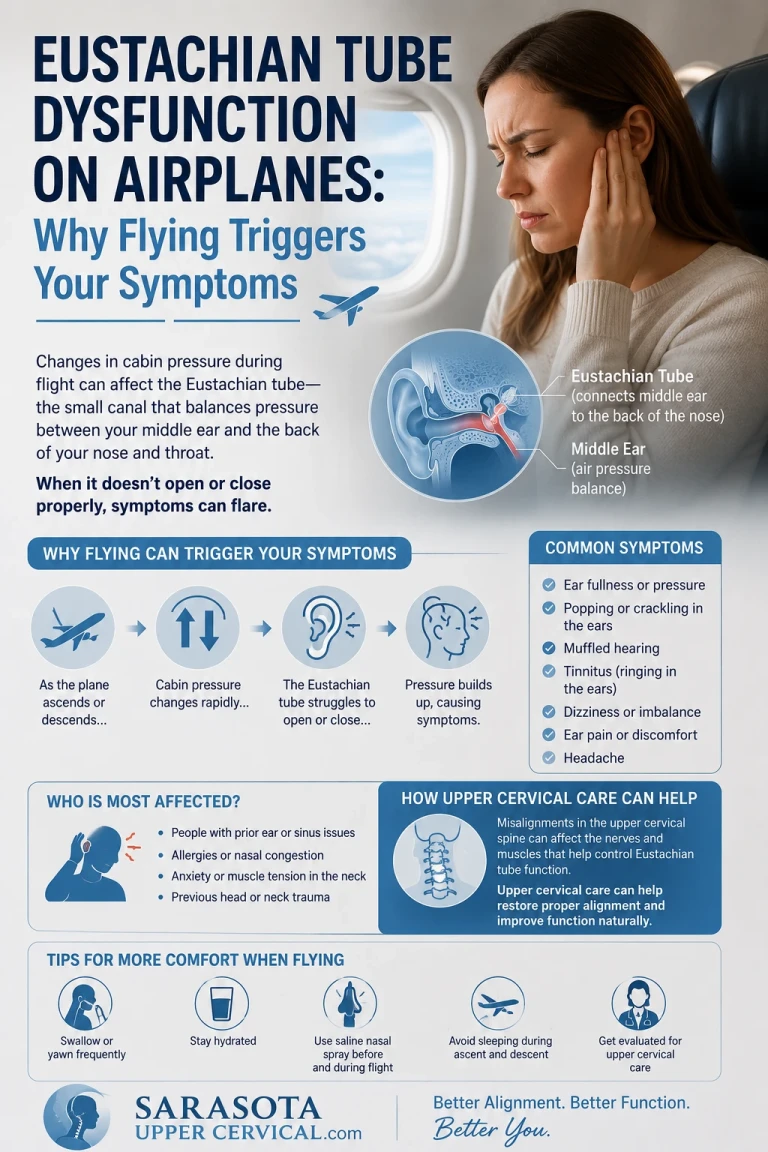
Eustachian Tube Dysfunction on Airplanes: Why Flying Triggers Your Symptoms
Jun 08, 2026Why Deep Sleep Specifically Takes the Hit
Patients often report something more particular than "I can't sleep." They report that they sleep seven hours and wake up as though they slept two. That pattern points at slow-wave sleep rather than total sleep time.
In a healthy night, the descent into deep non-REM sleep is accompanied by a pronounced autonomic shift: heart rate falls, blood pressure dips, and vagal tone rises. This nocturnal dip is one of the more robust markers of restorative sleep. In insomnia patients with objectively short sleep duration, that dip is blunted — the wake-to-sleep heart rate reduction is smaller than in good sleepers, and heart rate variability measures are reduced. This is established, though the literature is genuinely mixed and several studies have failed to find the autonomic differences, which is why I am describing it as a pattern rather than a universal finding.
If sympathetic tone stays elevated through the night, the physiological conditions for slow-wave sleep are never fully established. The architecture degrades even when the clock time in bed looks adequate. That is the mechanistic account of why a person can log eight hours and feel unrefreshed — and it is why total sleep time is a poor outcome measure in this population.
Objective Testing Before Anyone Touches Your Neck
This is the part of upper cervical practice I care about most, and it is the part most often skipped. A structural hypothesis about your atlas is worthless unless it is measured, and measured before and after.
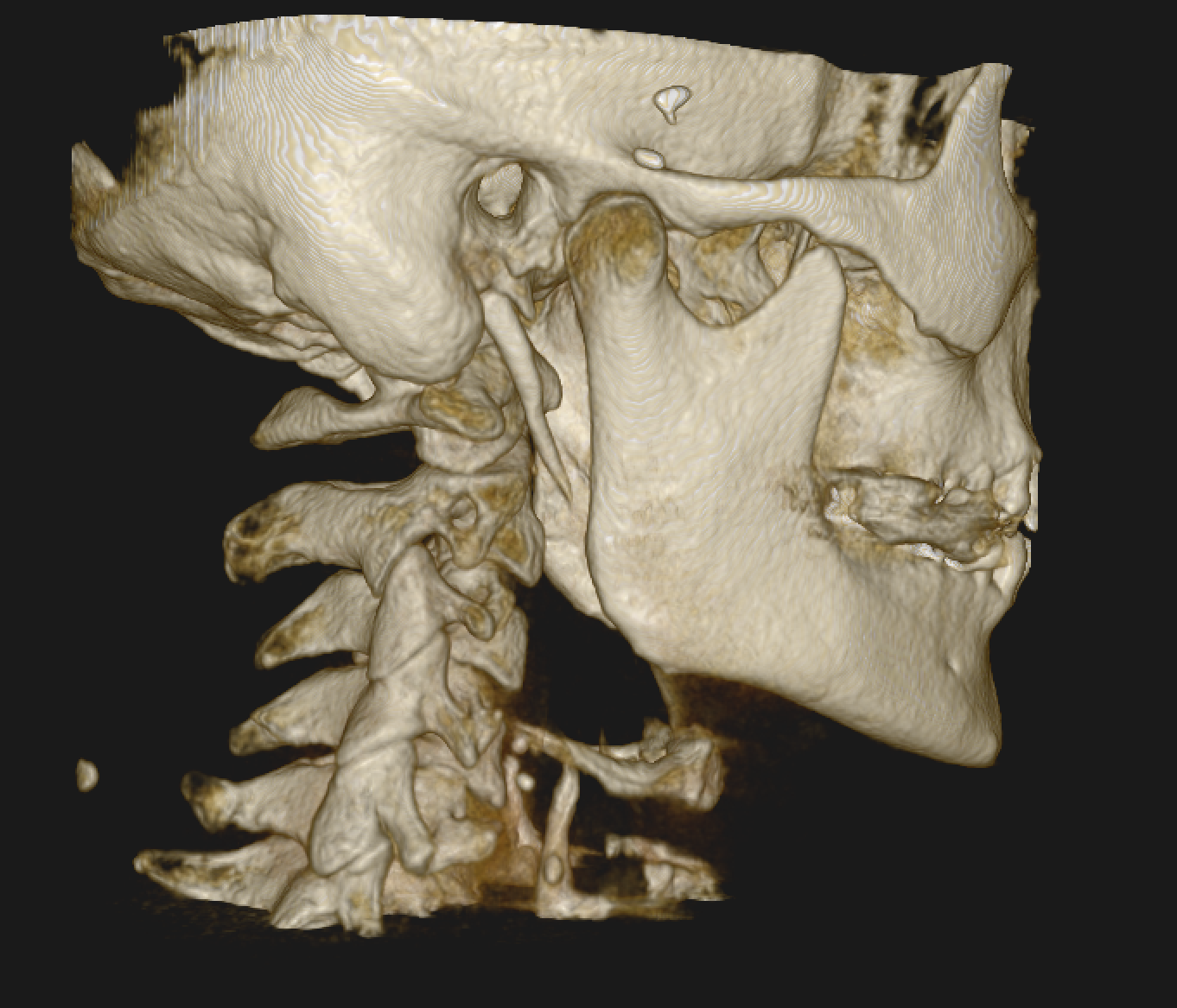
Cone beam computed tomography is the imaging standard we use for the craniocervical junction. Unlike a plain radiograph, which superimposes structures and forces the examiner to infer three-dimensional position from a two-dimensional shadow, CBCT reconstructs actual volumetric anatomy — the condyles, the atlas lateral masses, the odontoid, the atlanto-occipital and atlantoaxial articulations, and the surrounding neurovascular canals. A 2022 literature review examining CBCT for upper cervical chiropractic application concluded that visualisation of incidental and abnormal craniocervical junction findings was superior to radiography, and that effective radiation dose for comparable protocols was equal to or less than radiography. The honest qualifier is that CBCT dose varies substantially with field of view and protocol, published head-and-neck figures span a wide range, and that review appeared in the chiropractic literature rather than in mainstream neuroradiology. CBCT also shows bone, not nerve — it will not image irritation or inflammation the way MRI can, and it is not a substitute for MRI where soft tissue or cord pathology is a concern.
Alongside imaging, meaningful objective testing before and after correction should include some measure of autonomic state, and heart rate variability is the most accessible one — it is the same class of measurement the insomnia hyperarousal literature relies on. Paraspinal thermal scanning is used in much of the upper cervical field to assess autonomic asymmetry; I will be straightforward that thermographic assessment has a contested evidence base and I regard it as one input among several rather than a diagnostic standard. Postural and functional measures — head tilt, weight distribution asymmetry, cervical joint position error testing of the kind described by Revel and by Sterling — round out the picture. Actigraphy or a validated sleep diary matters too, because subjective sleep reporting is unreliable in exactly this population.
The principle underneath all of it is simple. If we cannot measure a change, we cannot claim one.
Correction Without Twisting, Popping, or Pulling
The image most people carry of chiropractic — the rotational thrust, the audible cavitation, the hands cradling the head and turning it sharply — is not what happens in an upper cervical correction of the type we perform.
There is no twisting of the neck. There is no forced rotation. There is no pulling or traction of the head. There is no cracking or popping sound, because no joint cavitation is being induced. The patient lies on their side on a specialised table, and a precisely calculated, low-force contact is applied to the atlas along a vector derived from that individual's imaging. The correction is measured in ounces of force, not pounds, and it is directed by the CBCT analysis rather than by feel.
Two reasons this matters. Practically, most people carrying a significant craniocervical injury history are apprehensive about having their neck manipulated, and that apprehension is not irrational. More substantively, if the working hypothesis is that disordered upper cervical afferent input is driving elevated sympathetic tone, then introducing a high-velocity rotational stimulus into an already-sensitised region is at odds with the goal. A low-force, image-guided, non-rotational correction is the approach that follows logically from the mechanism.
I should say clearly that the superiority of low-force upper cervical technique over other manual approaches for sleep outcomes specifically has not been established in comparative clinical trials. The rationale above is mechanistic reasoning, not trial evidence.
Related article
What This Approach Can and Cannot Claim
The evidence base for upper cervical chiropractic care and insomnia consists of case reports and small case series. There are no randomised controlled trials establishing efficacy for insomnia. Studies examining spinal manipulation and heart rate variability exist and some show short-term autonomic shifts, but they are small, methodologically heterogeneous, and do not demonstrate durable clinical sleep benefit.
So the honest statement is this. Upper cervical care is not a treatment for insomnia, and it is not a substitute for cognitive behavioural therapy for insomnia, which remains the best-evidenced first-line intervention and which I recommend without reservation. What upper cervical care addresses is a structural and neurological finding — atlas misalignment with associated altered afferent input — in patients where that finding is objectively demonstrated. Whether correcting it improves your sleep is an individual question answered by measurement over a defined trial period, not by a promise made in advance.
If you have had a documented craniocervical trauma, if your sleep problems began or worsened after it, if you have accompanying symptoms suggesting upper cervical involvement — headaches originating at the skull base, dizziness with head position change, unexplained tinnitus, unilateral symptom patterns, neck stiffness — and if conventional sleep treatment has genuinely failed, then this line of investigation is reasonable. If none of that describes you, it probably is not, and I would rather tell you that at the outset.
When Insomnia Is Not a Neck Problem
Several conditions produce sleep disruption and require entirely different management, and missing them because someone was fixated on the cervical spine would be a serious failure.
Obstructive sleep apnoea is the most important. If you snore, if a partner has witnessed breathing pauses, if you wake gasping, if you have significant daytime sleepiness, or if you carry the associated risk factors, you need a sleep study before anything else. Restless legs syndrome, periodic limb movement disorder, and narcolepsy likewise require sleep medicine evaluation. Thyroid dysfunction, perimenopause, uncontrolled pain, nocturia, and a long list of medications — including some antidepressants, stimulants, beta-blockers and corticosteroids — all disrupt sleep through mechanisms that have nothing to do with your atlas. Depression and anxiety disorders both cause and are caused by insomnia and warrant proper mental health assessment rather than a structural explanation.
And there are findings that mean you should be evaluated urgently rather than adjusted: severe sudden-onset headache unlike any you have had, progressive neurological deficit, weakness or numbness in the limbs, changes in bowel or bladder control, difficulty swallowing or speaking, unexplained weight loss, fever with neck stiffness, or any suspicion of craniocervical instability. Those are medical situations, and a responsible upper cervical practice refers them out rather than working on them.
Insomnia Evaluation in Sarasota, Bradenton, and Lakewood Ranch
If you are in Sarasota, Bradenton, or Lakewood Ranch, and your sleep problems trace back to a car accident, a sports injury, a fall, or a concussion that nobody ever connected to them, an upper cervical evaluation will at minimum tell you whether there is a structural finding to explain it. CBCT imaging, autonomic and functional testing, and a straightforward conversation about what the findings do and do not support — followed by a defined trial with measured outcomes if, and only if, the findings warrant it.
To learn more about upper cervical care or to schedule an evaluation call 941 259-1891.
Medical Disclaimer
This article is provided for general educational purposes only and does not constitute medical advice, diagnosis, or treatment. It does not establish a doctor-patient relationship. Upper cervical chiropractic care is not a treatment or cure for insomnia or any other named disease, and no guarantee of any particular result is made or implied. Individual results vary, and the mechanisms described above include both established physiology and hypotheses that have not been demonstrated in clinical trials, as indicated throughout the text. Always consult a qualified healthcare provider regarding any sleep disorder or medical condition, and do not discontinue or alter prescribed treatment without consulting the prescribing physician. In accordance with Florida Administrative Code Rule 64B2-15.001, this content is offered without any claim of superiority of one method of treatment over another and without any promise of cure or guaranteed outcome.
Written by Dr. Drew Hall, upper cervical chiropractor, Sarasota Upper Cervical / Hall Upper Cervical Chiropractic PC, serving Sarasota, Bradenton, and Lakewood Ranch, Florida.


Leave a comment