

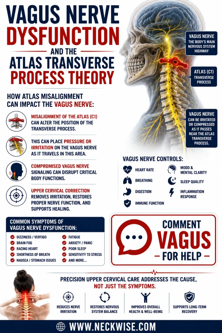
The vagus nerve is the longest cranial nerve in the body, carrying roughly eighty percent of its fibers as afferent signals traveling from the organs back up to the brainstem. It regulates heart rate variability, digestive motility, airway tone, and the parasympathetic braking system that keeps the sympathetic nervous system from running unchecked. Because it threads directly through the upper cervical spine on its way out of the skull, the vagus nerve is one of the few structures in the body where a purely mechanical problem in the neck can produce what looks, on the surface, like a purely medical one. This is the foundation of what we call the atlas transverse mechanical traction model: the idea that a loss of normal cervical curve and the resulting postural collapse around the C1 vertebra places sustained mechanical traction on the vagus nerve at the level of the atlas transverse process, and that this traction — not infection, not primary autoimmune disease, not a psychiatric diagnosis — is the upstream driver of a wide constellation of seemingly unrelated symptoms.
The Vagus Nerve's Vulnerable Passage Through the Upper Cervical Spine
To understand why the atlas matters so much to vagal function, it helps to trace the nerve's actual path. The vagus nerve exits the skull through the jugular foramen alongside the glossopharyngeal and spinal accessory nerves and the internal jugular vein. Immediately after leaving the skull, these structures are forced through a narrow corridor between the transverse process of C1 and the styloid process, with the facial nerve running nearby. From there, the vagus nerve drops into the carotid sheath, a fascial tube that also contains the internal carotid artery, the internal jugular vein, and the cervical sympathetic chain. The carotid sheath sits directly behind the sternocleidomastoid muscle, running from the base of the skull down to the first rib and sternum.
Schedule Your Appointment
Schedule appointmentThe geometry here is the crux of the problem. As the vagus nerve transitions from the jugular foramen down into the carotid sheath, it makes a sharp turn — close to ninety degrees — right at the atlas transverse process. Two of the vagus nerve's most important relay stations, the superior jugular ganglion and the inferior nodose ganglion, sit almost on top of the craniocervical junction, close enough that instability at C1 can produce direct structural strain on the nodose ganglion itself. A nerve that makes a sharp directional turn through a narrow bony-fascial corridor is, by definition, a nerve with very little slack. It does not tolerate being stretched, and it does not tolerate being compressed. Upper cervical instability puts it at risk of both.
The Atlas Transverse Mechanical Traction Model
The atlas transverse mechanical traction model describes the specific mechanical sequence that turns normal anatomy into a source of chronic dysfunction. When the atlas (C1) loses its correct positional relationship with the skull and with C2 — whether from a car accident, a fall, years of forward head posture from screen use, or cumulative ligament laxity — the vertebra does not simply sit still and become "stuck." It becomes unstable, meaning it moves more than it should, in directions it should not. As the head migrates forward relative to the shoulders and the cervical curve straightens or reverses, the soft tissue architecture around the atlas transverse process is placed under sustained tensile load. The scalenus anterior, which attaches to the anterior transverse processes, and the levator scapulae, which attaches along the lateral C1 through C4 transverse processes, tighten in response to forward head posture. Because the carotid sheath and its contents sit directly against this same muscular and osseous landscape, the vagus nerve is dragged along with it.
This is traction in the literal biomechanical sense: a nerve fixed at one end by the jugular foramen and constrained further down by the carotid sheath, subjected to a sustained pull as the vertebra above it shifts out of its normal architecture. Unlike an acute compression injury, this kind of traction tends to be low-grade, chronic, and cumulative — which is exactly why vagus nerve dysfunction of this type so often develops silently over years rather than announcing itself with a single dramatic event.
What the Research Shows
This is not a new idea invented to explain away hard-to-diagnose symptoms; it has a growing body of published support, much of it from Ross Hauser, MD, and colleagues at their Fort Myers research clinic. Hauser's group has described dynamic carotid sheath compression from ligamentous cervical instability as a mechanism that affects the vagus nerves and internal jugular veins, occurring primarily at the level of the atlas, and has proposed that a large share of body-wide symptoms and disease trace back to vagus nerve dysfunction or degeneration, while brain-specific symptoms trace back to internal jugular vein compression and the resulting rise in cerebrospinal fluid and venous pressure.
A 2025 paper from the same research group, published in Frontiers in Neurology and describing what the authors term cervicovagopathy, illustrates the anatomy directly: the vagus nerve runs close to the anterior cervical vertebrae, and this proximity is most pronounced at the atlanto-axial joint, where the nerve is described as highly vulnerable to traction and stretch. The same paper traces the mechanism back to forward head posture from prolonged computer and phone use, which produces cervical ligament laxity through a slow stretching process referred to in the biomechanics literature as "creep." As the cervical vertebrae subluxate anteriorly under this laxity, both the internal jugular veins and the autonomic nerves of the anterior neck, including the vagus nerve and the cervical sympathetic ganglion, are placed under stretch-compression.
A follow-up 2026 Frontiers study examining more than two hundred patients evaluated for cervical spine etiology reinforced this picture with objective imaging findings. The researchers found forward head posture, loss of cervical lordosis measured by depth of curve, and ligamentous instability in both the upper and lower cervical spine to be near-universal in their symptomatic cohort, alongside measurable internal jugular vein compression and reduced vagus nerve diameter suggestive of degeneration or atrophy. Their working model proposes two parallel mechanisms: jugular vein compression raising intracranial and cerebrospinal fluid pressure, and vagus nerve degeneration disrupting autonomic regulation, together producing what they describe as a wide range of brain-based and body-based symptoms.
Related article
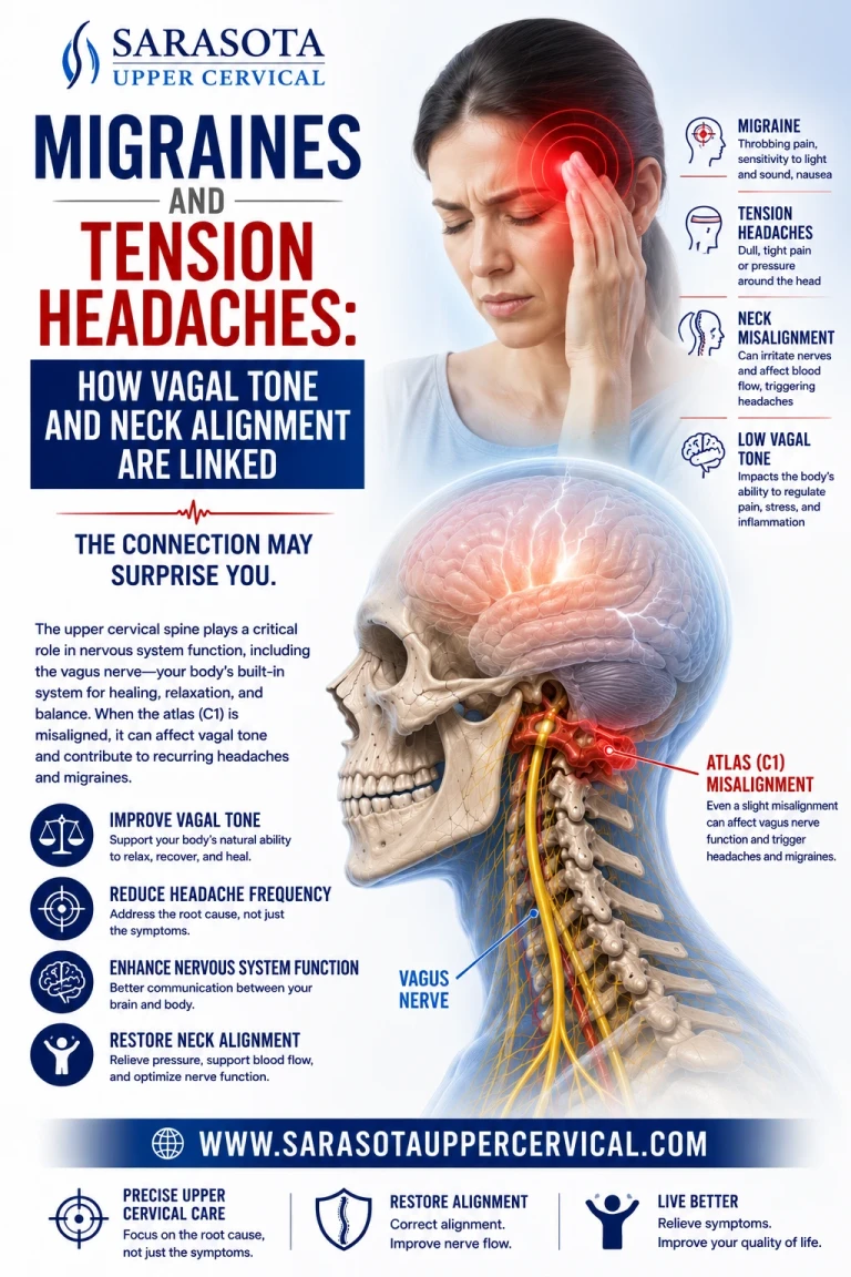
Independent research on cervical alignment adds further support from a different angle. Chiropractic BioPhysics case data has documented resolution of chronic migraine alongside objectively measured improvement in cervical lordosis, and separate radiographic research has linked improved cervical lordosis and reduced forward head posture to improved central conduction time on somatosensory-evoked potential testing — a direct neurophysiological measure that structural correction of the cervical curve translates into measurable nervous system change, not just symptomatic improvement that could be explained by other factors.
Loss of the Cervical Curve as the Mechanical Driver
The cervical curve is not a cosmetic feature of the spine; it is a load-distribution system. A properly maintained lordotic curve spreads the weight of the head across the entire cervical column and keeps the soft tissue structures that pass through it, including the carotid sheath and the vagus nerve, in a position of relative slack. When that curve straightens or reverses, several things happen simultaneously. The sagittal vertical axis increases as the head translates forward of the shoulder plane, forward head posture becomes structurally locked in rather than merely postural, and the muscles that attach along the C1 through C4 transverse processes shorten and tighten to keep the head from falling forward entirely.
Because the vagus nerve, the internal jugular vein, and the cervical sympathetic chain all travel through this same narrow anterior corridor, none of them get a pass when the curve collapses. The loss of cervical curve is, in this model, the mechanical event that converts a normally slack, well-protected vagus nerve into one under continuous low-grade tension at the atlas transverse process. This is also why the atlas transverse mechanical traction model treats loss of cervical curve not as a downstream consequence of neck pain but as a primary driver of autonomic symptoms that can appear in patients who do not report significant neck pain at all.
The Downstream Effects of Vagus Nerve Dysfunction
Because the vagus nerve regulates so much of the body's autonomic and organ-level function, its dysfunction rarely produces a single, clean symptom. Patients with vagus nerve involvement of this type commonly present with cardiovascular and autonomic complaints such as postural orthostatic tachycardia syndrome and broader dysautonomia, marked by an unstable heart rate, blood pressure swings, and poor tolerance for standing or exertion. Digestive motility is frequently affected, since the vagus nerve is the primary parasympathetic supply to the stomach and intestines, producing bloating, delayed gastric emptying, and irregular bowel function. Because the same jugular foramen crowding and upper cervical instability that stretches the vagus nerve also affects the nearby brainstem nuclei that govern sympathetic outflow — including the nucleus tractus solitarius, the caudal ventrolateral medulla, and the rostral ventrolateral medulla — patients often develop symptoms of sympathetic disinhibition layered on top of vagal withdrawal, which shows up clinically as anxiety, migraine and tension-type headache, and tinnitus. Immune and inflammatory regulation, which the vagus nerve influences through the cholinergic anti-inflammatory pathway, can also be affected, and this mechanism has been proposed as a contributing factor in conditions such as rheumatoid arthritis and in the persistent autonomic symptoms reported after Long COVID infection.

Why Precision Imaging Changes the Correction
If the atlas transverse mechanical traction model is correct, then the correction has to address the actual three-dimensional position of the atlas relative to the skull and axis, not simply loosen the surrounding muscles or apply generic spinal manipulation. This is the reasoning behind the use of cone beam computed tomography, or CBCT, in upper cervical care. CBCT produces a true three-dimensional reconstruction of the atlas and axis, showing the exact orientation of the joint surfaces, the individual angles at which they sit, and any compensation patterns elsewhere in the cervical spine, at a level of detail that cannot be felt by hand or seen on a standard two-dimensional film. Because no two atlas joints are shaped or angled the same way, this imaging allows the correction to be calculated for the individual patient's own anatomy rather than applied as a one-size-fits-all technique.
Just as importantly, the correction itself looks nothing like general chiropractic manipulation. Upper cervical procedures such as Blair are built specifically to avoid twisting, popping, cracking, or pulling the neck. Blair and Knee Chest approaches use the patient's own anatomy from CBCT imaging to calculate a precise, low-force, non-rotational correction, often delivered with a small, specific thrust. . In every case, the goal is the same: restore the correct three-dimensional relationship of the atlas to the skull and axis without ever placing torque or traction on the very structures — the vagus nerve, the internal jugular vein, the cervical sympathetic chain — that the correction is meant to relieve pressure from in the first place. Follow-up imaging is then used to confirm the correction has held, turning the entire process into a measurable, repeatable structural intervention rather than a subjective one.
Restoring the Curve, Restoring Vagal Function
Taken together, the anatomy of the jugular foramen and carotid sheath, the biomechanics of forward head posture and cervical curve loss, and the growing body of research out of Fort Myers and elsewhere point toward the same conclusion: a straightened or reversed cervical curve is not a benign finding on an X-ray. It is a structural event capable of placing the vagus nerve under sustained traction at the atlas transverse process, with downstream consequences for the cardiovascular, digestive, and nervous systems. Correcting that traction requires seeing the atlas in three dimensions and correcting its position with the same precision used to identify the problem in the first place, without ever resorting to the twisting, popping, or pulling that put the nerve at risk to begin with.
This article is for educational purposes and is not intended as a substitute for individualized medical or chiropractic evaluation. The mechanisms described reflect an emerging area of clinical research, and evidence in this field continues to develop. Anyone experiencing symptoms of autonomic or vagus nerve dysfunction should be evaluated by a qualified healthcare provider.
Dr. Drew hall For a structural evaluation of your cervical curve and upper cervical alignment


Leave a comment